The objective of the current study was to investigate the role of the MDH enzyme system in liver cells of alloxan-induced diabetic rats and to examine how the administration of inulin from Jerusalem artichoke affects the enzyme’s activity and gene transcription in these diabetic rats’ livers. In this study, male Wistar rats weighing 150-200 g were selected for the experiment. Diabetes mellitus was induced by a single intraperitoneal injection of 5% alloxan monohydrate (in 0.9% saline). The control group received an equivalent amount of saline solution. Statistical analysis was performed using StatTech v. 1.2.0 software. An increase in NAD-dependent malate dehydrogenase (MDH) activity, accompanied by the emergence of a novel liver isoform, was observed in rats with alloxan-induced diabetes. This finding suggests the potential involvement of the malate dehydrogenase enzyme system in the body’s adaptive response to oxidative stress induced by biochemical changes in diabetic cells. In type I diabetes, this rise in enzyme activity is associated with the appearance of an additional MDH isoform in peroxisomes. Gene expression analysis of mdh1 and mdh2 indicates that diabetes triggers enzyme activation at the gene transcription level. When inulin was administered, it significantly reduced blood glucose levels in rats with alloxan-induced diabetes and restored the regular transcriptional activity of these genes. Consequently, the formation of the new MDH isoform was prevented. This suggests that inulin could be a promising option for pharmacologically managing the metabolic changes associated with diabetes-related pathologies.
Diabetes mellitus (DM) has become one of the most prevalent chronic conditions worldwide, with the number of affected individuals continually on the rise [1, 2]. The International Diabetes Federation (IDF) estimates that by 2040, the global population diagnosed with diabetes will surpass 600 million people [3].
Among the numerous protective strategies against diabetes-induced cellular damage, natural compounds derived from plant extracts hold significant promise [4, 5]. For instance, Jerusalem artichoke (Helianthus tuberosus) leaves and tubers, which are rich in inulin, contain substantial amounts of fructans, soluble dietary fibers, sesquiterpenes, diterpenes, and chlorogenic acid analogs [6, 7]. Inulin and other bioactive components in Jerusalem artichoke concentrate have been shown to influence pancreatic islet development, particularly during the first 3 weeks of administration, thereby alleviating the severity of alloxan-induced diabetes in rats [8, 9]. Furthermore, inulin can bind glucose, improving the responsiveness of insulin-sensitive cells and ultimately lowering blood glucose levels [10].
The process by which the body adapts to alloxan-induced diabetes in animal models is intricate and involves several stages, with metabolic shifts at the cellular level being a key component [11, 12]. The activation of enzymes within the glyoxylate cycle and the tricarboxylic acid cycle leads to changes in fundamental metabolic pathways, including glycogen resynthesis in the liver of rats with experimental diabetes and conditions associated with nutritional deprivation [13, 14]. In this context, gluconeogenesis plays a central role in the body’s adaptive response, while the Krebs cycle’s involvement in energy metabolism is equally critical [15, 16]. As a result, the body’s adaptation hinges on the balance between glucose breakdown and synthesis within the liver and other tissues. A central enzyme system that contributes to these adaptive processes is malate dehydrogenase (MDH) [17]. This enzyme complex plays a vital role in maintaining energy and metabolic pathways, functioning across various physiological processes [18].
In light of this, the objective of the current study was to investigate the role of the MDH enzyme system in liver cells of alloxan-induced diabetic rats and to examine how the administration of inulin from Jerusalem artichoke affects the enzyme’s activity and gene transcription in these diabetic rats’ livers.
In this study, male Wistar rats weighing 150-200 g were selected for the experiment. The rats were housed in a controlled environment with natural light cycles, with unrestricted access to water and food.
Diabetes mellitus was induced by a single intraperitoneal injection of 5% alloxan monohydrate (in 0.9% saline). The control group received an equivalent amount of saline solution.
After the rats developed diabetic symptoms, they were randomly assigned to three groups: “Norm” (control), “Diabetes” (diabetic), and “Diabetes + INULIN” (inulin-treated). The “Diabetes + INULIN” group received a daily dose of 60 mg/kg of inulin for 14 days, while the “Norm” and “Diabetes” groups were given distilled water.
The aqueous extract of Jerusalem artichoke leaves (Helianthus tuberosus) was prepared by grinding dry leaves into powder, which was then steeped in hot water at a ratio of 5 g per 100 ml for 24 hours. The solution was filtered, and the resulting extract was added to drinking water at a dosage of 60 mg/kg/day [19].
Liver samples were collected by decapitating the animals under ether anesthesia [20, 21]. Blood glucose levels were measured using a “Satellite Plus” glucose meter, and blood samples were drawn from the tail vein after a 12-hour fasting period.
To prepare liver tissue homogenates, the liver was homogenized in a cold Tris-HCl buffer (50 mM, pH 7.8) containing EDTA, MgCl2, and DTT, and centrifuged at 5000 g for 10 minutes. The supernatant was collected for subsequent analysis.
The activity of malate dehydrogenase (MDH) was measured by spectrophotometry on an SF-2000 spectrophotometer at 340 nm, monitoring NADH consumption. The assay mixture contained tris-HCl buffer (50 mM, pH 7.5), oxaloacetate (1.5 mM), NADH (0.15 mM), MgCl2 (5 mM), and DTT (4 mM). MDH activity was quantified as enzyme units per mg of protein, where one unit corresponds to the conversion of 1 micromole of substrate per minute at 25 °C.
To analyze MDH isoenzymes, electrophoresis was performed on polyacrylamide gels prepared with 7.5% separating and 2% stacking gels, according to the modified Davis method [22]. The gels were subjected to electrophoresis in a tris-glycine buffer (0.05 M, pH = 8.6) with 0.01% bromophenol blue as a tracking dye. MDH activity was visualized by the tetrazolium staining method, which included the following reagents: tris-HCl buffer (50 mM, pH = 7.5), sodium malate (0.2 M), NAD+ (3 mM), MgCl2 (1 mM), phenazine metasulfate (0.01 M), and nitroblue tetrazolium (0.01 M). A total of 5 µg of protein was loaded per gel pocket. Protease inhibitors (0.1 mM n-chloromercury benzoate and 0.1 mM phenylmethylsulfonyl fluoride) were added to prevent protein degradation.
To determine the subcellular localization of MDH, an isodensity gradient centrifugation was performed using sucrose gradients on a Beckman centrifuge. The gradient was composed of sucrose solutions at concentrations ranging from 2.5 M to 1.3 M in a 50 mM Tris-HCl buffer (pH 7.5) containing EDTA, DTT, and other components. Fractions were collected, diluted to 0.4-0.5 M sucrose, and centrifuged at 12,000 g for 30 minutes. The organelles were then analyzed using marker enzymes for cytoplasm (alcohol dehydrogenase), mitochondria (succinate dehydrogenase), and peroxisomes (catalase).
Total RNA was extracted from liver tissues using the phenol-chloroform method, followed by LiCl precipitation [23, 24]. RNA integrity was assessed by agarose gel electrophoresis stained with ethidium bromide.
For cDNA synthesis, mRNA was reverse-transcribed using M-MuLV reverse transcriptase and oligo dT primers according to the manufacturer’s protocol. The primers for the mdh1 and mdh2 genes were selected based on their nucleotide sequences available in the NCBI database (https://www.ncbi.nlm.nih.gov/genes), using the Primer-BLAST tool. The mdh1 primers were: forward—5-gctctactcgttccctgtcg-3, reverse—5-acgactgtgtagtcatgcgg-3, and the mdh2 primers: forward—5-acccccaaggttgactttcc-3, reverse—5-ttccttcccattcatggcgt-3.
Polymerase chain reaction (PCR) amplification was performed using the AmpliSence reagent set (Helicon, Russia) on a LightCycler 96 system (Roche, Switzerland). The thermal cycling conditions included an initial denaturation at 95 °C for 5 minutes, followed by 40 cycles of denaturation at 95 °C for 20 seconds, annealing at 60 °C for 30 seconds, and extension at 72 °C for 40 seconds. A final elongation step was performed at 72 °C for 10 minutes.
Statistical analysis was performed using StatTech v. 1.2.0 software. The normality of data distributions was assessed using the Shapiro-Wilk test (for samples < 50) or Kolmogorov-Smirnov test (for samples ≥ 50) [25]. Quantitative data were presented as means ± standard deviation (SD) with 95% confidence intervals (CI). Group comparisons were performed using one-way ANOVA, and post-hoc Tukey’s test was used for pairwise comparisons when variances were equal. Statistically significant differences were considered at P < 0.05.
Before alloxan administration, blood glucose levels across all experimental groups were nearly equal (P = 0.006). However, after 72 hours of alloxan exposure, glucose concentrations were significantly elevated in the experimental groups, with the “Diabetes” group measuring 19.3 mmol/L and the “Diabetes+INULIN” group at 19.72 mmol/L (Figure 1). The “Norm” and “Diabetes” groups received distilled water, while the “Diabetes+INULIN” group was treated with a daily dose of inulin solution (60 mg/kg body weight) for fourteen days. At the conclusion of the study period, glucose levels in the “Diabetes+INULIN” group decreased markedly compared with the “Diabetes” group, from 19.95 mmol/L to 8.95 mmol/L. The control group, consisting of healthy rats, consistently maintained blood glucose levels within the normal range throughout the experiment, averaging 4.8-5.4 mmol/L. The observed reduction in hyperglycemia in the “Diabetes+INULIN” group may be attributed to the Jerusalem artichoke leaf extract’s potential to block glucose absorption in the intestines, promoting glucose release from the liver [26, 27]. Additionally, the inulin component of the extract may stimulate pancreatic insulin production [16].
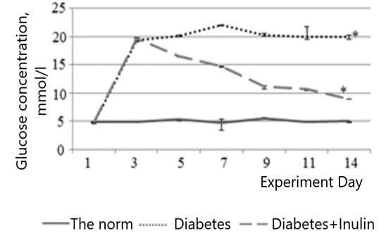
Figure 1. Changes in blood glucose levels over time in the different rat groups. The “Norm” group consists of control rats (n = 12), the “Diabetes” group includes rats induced with alloxan diabetes (n = 12), and the “Diabetes+INULIN” group includes diabetic rats treated with inulin solution (n = 12).
The comparison of glucose concentrations between the Diabetes and Diabetes+INULIN groups throughout the experiment revealed significant differences (P = 0.007 for p11day–1day = 0.009, P = 0.006 for p3day–1day = 0.009). In contrast, no such differences were observed in the Norm group (P = 0.207) by the Kruskal-Wallis test.
Regarding MDH activity in rat liver hepatocytes, two separate pools of enzyme activity were identified in healthy rats: cytoplasmic and mitochondrial. However, in diabetic rats, MDH activity was also detected in the peroxisomal fraction (Table 1). The cytoplasmic and mitochondrial forms in the control group accounted for 46.0% and 54.0% of the total, respectively. In diabetic rats, the distribution was altered, with 25.0% in the cytoplasm, 58.0% in the mitochondria, and 16.0% in the peroxisomes. The cross-contamination level was determined to be 4-6%, which is within acceptable limits for interpreting the results.
Table 1. Specific malate dehydrogenase activity in different subcellular compartments of liver hepatocytes in control rats (Norm), rats with alloxan-induced diabetes (Diabetes), and diabetic rats treated with inulin (Diabetes + INULIN)
Indicator | The norm | Diabetes | Diabetes + INULIN |
MDH activity in the cytoplasm, Units/mg of protein; M ± SD (95% CI) | 1.4 ± 0.27* (1.13-1.67) | 2.12 ± 0.15** (1.95-2.29) | 1.57 ± 0.27** (1.26-1.87) |
MDH activity in mitochondria, Units/mg of protein; M ± SD (95% CI) | 1.5 ± 0.19*** (1.3-1.69) | 4.62 ± 0.1* (4.51-4.74) | 1.85 ± 0.1*** (1.73-1.96) |
MDH activity in peroxisomes, Units/mg of protein; M ± SD (95% CI) | — | 0.81 ± 0.13 (0.66-0.96) | — |
Note. According to the results, a statistically significant difference in MDH activity was identified between cytoplasmic and mitochondrial levels (*P < 0.001; **P Diabetes–Diabetes+INULIN = 0.003; ***P NORM–Diabetes+INULIN = 0.027*). Method used: Variance analysis.
In animals with DM treated with INULIN, no MDH activity was detected in the peroxisomal fraction, and the MDH activity in the cytoplasmic and mitochondrial fractions was nearly identical to the normal values (44% and 56%, respectively). A similar outcome was observed in a study in which an aqueous extract of European Olive (Olea europaea) was administered to rats with DM, demonstrating that the extract helps restore MDH activity to baseline levels [28].
The increase in MDH activity suggests that specific metabolic pathways involving this enzyme might be more active. However, the exact reasons for this rise in MDH activity are not entirely understood and require further investigation. Interestingly, other studies have indicated a similar shift in the peroxisomal distribution of MDH activity in rat hepatocytes during fasting conditions [29].
To investigate the activation of metabolic pathways linked to MDH activity in alloxan-induced diabetes, the MDH isoenzyme profile was examined. Electrophoresis using polyacrylamide gel revealed that, unlike control rats and those treated with plant extracts, which displayed two isoforms—MDH1 (cytoplasmic) and MDH2 (mitochondrial) with Rf values of 0.29 and 0.36, respectively—an additional MDH isoform, MDH3, was detected in rats with diabetes. This newly induced isoform migrated more slowly on the gel, with an Rf value of 0.20, and was found in the peroxisomal fraction (Figure 2).
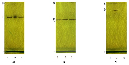
Figure 2. Distribution of malate dehydrogenase isoenzymes in liver fractions: cytoplasmic (a), mitochondrial (b), and peroxisomal (c) from control rats (1), rats with alloxan-induced diabetes (2), and diabetic rats treated with inulin (3). S represents the starting point, P1, P2, and P3 denote different isoforms of malate dehydrogenase, and F indicates the position of the bromophenol blue dye marker front.
The data suggest that an increase in MDH activity is associated with the emergence of a new isoform localized to the peroxisomal fraction. Previous research has demonstrated that, in models of experimental diabetes and food deprivation, the activities of enzymes associated with the glyoxylate cycle, such as isocitrate lyase and malate synthase, are elevated in the kidneys and liver of rats [30]. Based on these findings, it is proposed that the inducible MDH isoform may be involved in gluconeogenesis. Moreover, inulin treatment in diabetic rats blocks the formation of the peroxisomal MDH.
The shift in isoenzyme distribution in alloxan-induced diabetic rats may result from altered expression of MDH-related genes. PCR-RV analysis shown in Figure 3 reveals that the transcript levels of these genes were notably higher in the livers of diabetic rats compared to control rats. Specifically, the mdh2 gene saw a more than twofold increase in transcription in diabetic rats, correlating with heightened mitochondrial MDH activity. Similarly, transcription of the mdh1 gene was elevated 1.8-fold in diabetic rats, which may be linked to the generation of an additional peroxisomal MDH isoform, potentially via alternative splicing. According to Stibler et al., a C-terminally expanded isoform containing the peroxisomal targeting signal 1 (PTS1) is generated by utilizing an alternative stop codon, which then targets the isoform to peroxisomes.
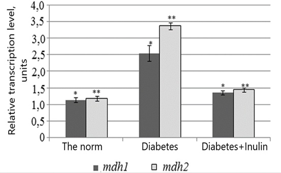
Figure 3. Comparison of transcript levels for the mdh1 (cytoplasmic) and mdh2 (mitochondrial) malate dehydrogenase genes in the liver of control rats (Norm), rats with alloxan-induced diabetes (Diabetes), and diabetic rats treated with inulin (Diabetes+INULIN). Statistical analysis of the transcription levels of the mdh1 and mdh2 genes across all groups showed significant differences (*P = 0.039; **P < 0.001). Analysis method: Variance analysis.
In conclusion, our findings show that experimental diabetes in rats leads to increased MDH activity and the development of a peroxisomal MDH isoform in the liver. The expression patterns of the mdh1 and mdh2 genes suggest that the enhanced enzyme activity is linked to increased transcription in diabetic conditions.
Additionally, the plant extract studied demonstrated hypoglycemic effects, as evidenced by a marked reduction in blood glucose levels in alloxan-induced diabetic rats. This treatment also counteracted the transcriptional changes in the studied genes and inhibited the formation of the peroxisomal MDH isoform in the diabetic rats.
We would like to thank our colleagues from the North Ossetian State Medical Academy for their valuable assistance during the experiment.
None
None
None
Open Access The author(s) retain copyright. This article is licensed under the Creative Commons Attribution-NonCommercial-ShareAlike 4.0 International License. It may be shared and adapted for non-commercial purposes with appropriate attribution, an indication of changes, and distribution of adaptations under the same license. Third-party material may be subject to separate terms identified in its credit line. View the license at https://creativecommons.org/licenses/by-nc-sa/4.0/.
 Publication System