Some studies have highlighted the role of Janus kinase 3 (JAK3) in the development of various cancers. To manage this condition, inhibitors such as decernotinib and facitinib are commonly used, although these drugs can cause elevations in liver enzymes and increased lipid levels. It is essential to recognize that new therapies are being developed to inhibit cancer cell growth, using both theoretical and experimental approaches. This study aimed to investigate whether carbazole derivatives (1-25) could interact with JAK3, using the 3pjc protein, decernotinib, and facitinib as reference compounds in the DockingServer tool. The results showed that the carbazole analogs engage with different regions of the 3pjc protein compared to facitinib and decernotinib. Additionally, the inhibition constant (Ki) for carbazole-protein interactions with compounds 2, 5, 9, 17, 18, and 22 was lower than that of the reference drugs, suggesting that these carbazole analogs could be effective JAK3 inhibitors and may help reduce cancer cell growth.
Cancer incidence has risen significantly across the globe in recent years, leading to a decrease in overall life expectancy [1-4]. Multiple factors have been identified as contributors to cancer development, including alcoholism [5], obesity [6, 7], smoking [8], and high-fat diets [9, 10]. Additionally, genetic factors are thought to play a role in the onset of various cancers [11-16]. For instance, mutations in the KRAS gene (Kirsten rat sarcoma virus) are known to promote the growth of cancer cells [17]. A study involving 436 French cancer patients found that 348 carried harmful mutations in the MSH2 tumor suppressor gene [18]. Moreover, mutations in the BCR-ABL gene, which encodes a tyrosine kinase, are associated with the development of leukemia [19]. Similarly, mutations in HER2, a membrane tyrosine kinase, have been linked to the development of breast cancer [20, 21]. Furthermore, cMYC overexpression, which is involved in regulating transcription factors, has been identified in breast cancer [22]. Mutations in the EGFR gene also contribute to lung cancer, with 15-20% of cases harboring such mutations, including 10% involving kinase domain insertions in exon 20 [23].
Conversely, emerging evidence suggests that the Janus kinase (JAK) family of cytoplasmic tyrosine kinases may influence cancer cell proliferation [24]. Specifically, JAK3 has been implicated in the growth of epithelial cancer cells [25]. Another research highlighted the role of the JAK3/STAT5 signaling pathway in gastric carcinoma progression [26]. Additionally, a mutation in the JAK3 gene (p.V715I) has been observed in both gastric and breast cancers [27]. Other research indicates that the JAK3/ERK pathway is involved in MMP-9 expression, which is linked to breast cancer cell growth [28]. These findings suggest that specific JAK3 gene mutations might be associated with various cancer types. In treatment, certain drugs are used to inhibit the growth of particular cancer cells. For example, PRN371, a JAK-3 inhibitor, has been shown to induce apoptosis in T cell lymphoma by disrupting the JAK3-STAT pathway [29]. These studies indicate that specific drugs can alter JAK-3's biological activity; however, the exact mechanism of action remains unclear. Thus, this study aims to investigate the binding of 26 carbazole compounds (Figure 1) to JAK3 using the 3pjc protein structure, with decernotinib (a JAK-3 antagonist) [30] and tofacitinib (a non-selective JAK-1 and JAK-3 antagonist) [31] as theoretical tools in the DockingServer program.
The structure of 26 carbazole derivatives is shown in Figure 1. These compounds were examined to determine their potential interactions with the JAK3 surface in various manners:
![Figure 1. Chemical structure of carbazole analogs (1-26): 1 = 2-(6-chloro-9H-carbazol-2-yl)propanoic acid; 2 = 5,21-dichloro-3-[(2R,3R,4R,5S,6R)-3,4-dihydroxy-6-(hydroxymethyl)-5-methoxyoxan-2-yl]-3,13,23-triazahexacyclo[14.7.0.02,10.04,9.011,15.017, 22]tricosa-1,4(9),5,7,10,15,17 (22),18,20-nonaene-12,14-dione; 3 = 1,2,3,4-Tetrahydrocarbazole; 4 = 1,2,7,8-Dibenzcarbazole; 5 = 1,5-Dimethyl-6H-pyrido(4,3-b)carbazole; 6 = 10-methoxy-7H-pyrido[4,3-c]carbazole; 7 = 10-Methoxy-7H-pyrido(2,3-c)carbazole; 8 = 11,12-Dihydro-6-methoxy-11-methylindolo(2,3-a)carbazole-5-carbonitri- le; 9 = 6,20-dihydroxy-13-[[2-hydroxy-1-(hydroxymethyl)ethyl]amino]-3-[3,4, 5-trihydroxy-6-(hydroxymethyl)tetrahydropyran-2-yl]-3,13,23-triazahexacy-clo[14.7.0.02,10.04,9.011,15.017,22]tricosa-1(16),2(10),4,6,8,11(15),17(22),18,20-nonaene-12,14-dione; 10 = 12-(2-Cyanoethyl)-6,7,12,13-tetrahydro-13-methyl-5-oxo-5H-indolo- (2,3-a)pyrrolo(3, 4-c)-carbazole; 11 = 3-ethyl-3,13,23-triazahexacyclo[14.7.0.02,10.04,9.011,15.017,22]trico-sa-1,4,6,8,10,15,17,19,21-nona-ene-12,14-dione; 12 = 13-(3-dimethylamino-2-hydroxypropyl)-3-methoxy-12-methyl-6,7,12, 13-tetrahydro-5H-indolo[2,3-a]pyrrolo[3,4-c]carbazole-5,7-dione; 13 = 3,4,5,6-Dibenzcarbazole; 14 = 5,11-Dimethyl-6H-pyrido(4,3-b)carbazole 2-oxide; 15 = 5,11-Dimethyl-6H-pyrido(4,3-b)carbazole; 16 = 5,7,9-Trimethyl-7H-dibenzo(c,g)carbazole; 17 = 5-(Hydroxymethyl)-11-methyl-6H-pyrido(4,3-b)carbazole; 18 = 5H-Naphtho[2,3-c]carbazole; 19 = 7-Hydroxymethyldibenzo(c,g)carbazole; 20 = 7H-Pyrido(4,3-c)carbazole; 21 = 9-(.beta.-Chloroethyl)carbazole; 22 = 9-Benzyl-9H-carbazole; 23 = 9-Ethyl-9H-carbazole; 24 = 9-Methyl-3-nitro-9H-carbazole; 25 = 9-[3-(3,5-Dimethyl-piperazin-1-yl)-propyl]-9H-carbazole; 26 = 4H-Benzo(def)carbazole (Source: https://pubchem.ncbi.nlm.nih.gob)](https://pubsys.eshragh.co/storage/files/pub-article/3c525205-8175-4689-ae43-ca66f84e5caa-XWhFhn2X2drIOXrS/image.jpg)
Figure 1. Chemical structure of carbazole analogs (1-26): 1 = 2-(6-chloro-9H-carbazol-2-yl)propanoic acid; 2 = 5,21-dichloro-3-[(2R,3R,4R,5S,6R)-3,4-dihydroxy-6-(hydroxymethyl)-5-methoxyoxan-2-yl]-3,13,23-triazahexacyclo[14.7.0.02,10.04,9.011,15.017, 22]tricosa-1,4(9),5,7,10,15,17 (22),18,20-nonaene-12,14-dione; 3 = 1,2,3,4-Tetrahydrocarbazole; 4 = 1,2,7,8-Dibenzcarbazole; 5 = 1,5-Dimethyl-6H-pyrido(4,3-b)carbazole; 6 = 10-methoxy-7H-pyrido[4,3-c]carbazole; 7 = 10-Methoxy-7H-pyrido(2,3-c)carbazole; 8 = 11,12-Dihydro-6-methoxy-11-methylindolo(2,3-a)carbazole-5-carbonitri- le; 9 = 6,20-dihydroxy-13-[[2-hydroxy-1-(hydroxymethyl)ethyl]amino]-3-[3,4, 5-trihydroxy-6-(hydroxymethyl)tetrahydropyran-2-yl]-3,13,23-triazahexacy-clo[14.7.0.02,10.04,9.011,15.017,22]tricosa-1(16),2(10),4,6,8,11(15),17(22),18,20-nonaene-12,14-dione; 10 = 12-(2-Cyanoethyl)-6,7,12,13-tetrahydro-13-methyl-5-oxo-5H-indolo- (2,3-a)pyrrolo(3, 4-c)-carbazole; 11 = 3-ethyl-3,13,23-triazahexacyclo[14.7.0.02,10.04,9.011,15.017,22]trico-sa-1,4,6,8,10,15,17,19,21-nona-ene-12,14-dione; 12 = 13-(3-dimethylamino-2-hydroxypropyl)-3-methoxy-12-methyl-6,7,12, 13-tetrahydro-5H-indolo[2,3-a]pyrrolo[3,4-c]carbazole-5,7-dione; 13 = 3,4,5,6-Dibenzcarbazole; 14 = 5,11-Dimethyl-6H-pyrido(4,3-b)carbazole 2-oxide; 15 = 5,11-Dimethyl-6H-pyrido(4,3-b)carbazole; 16 = 5,7,9-Trimethyl-7H-dibenzo(c,g)carbazole; 17 = 5-(Hydroxymethyl)-11-methyl-6H-pyrido(4,3-b)carbazole; 18 = 5H-Naphtho[2,3-c]carbazole; 19 = 7-Hydroxymethyldibenzo(c,g)carbazole; 20 = 7H-Pyrido(4,3-c)carbazole; 21 = 9-(.beta.-Chloroethyl)carbazole; 22 = 9-Benzyl-9H-carbazole; 23 = 9-Ethyl-9H-carbazole; 24 = 9-Methyl-3-nitro-9H-carbazole; 25 = 9-[3-(3,5-Dimethyl-piperazin-1-yl)-propyl]-9H-carbazole; 26 = 4H-Benzo(def)carbazole (Source: https://pubchem.ncbi.nlm.nih.gob)
The binding of the 26 carbazole derivatives to JAK3 was examined using the 3pjc protein structure [32], with decernotinib and facitinib used as reference controls in the DockingServer program [33].
The pharmacokinetic profiles of selected carbazole analogs (2, 5, 9, 17, 18, and 22) were analyzed using the SwissADME tool [34].
To evaluate the toxicity risks of carbazole analogs (2, 5, 9, 17, 18, and 22), as well as facitinib and decernotinib, the GUSAR software was employed, considering different routes of administration [35].
Previous studies suggest that certain carbazole derivatives might influence cancer cell development. For example, the drug alectinib has been utilized as a monotherapy for treating lung cancer [36]. Additionally, research indicated that 3,6-di(2-furyl)-9H-carbazole could act as a carcinogen by inhibiting topoisomerase II in MCF-7 breast cancer cells [37]. Another study demonstrated that specific N-acylated carbazole derivatives could reduce the growth of breast cancer cells using the MDA-MB-231 cell line [38]. Moreover, a tetrahydrocarbazole derivative was found to exert anticancer effects against lung cancer cells in a Calu-1 cell model [39].
Furthermore, the compound MHY407 has shown potential biological activity in treating breast cancer using MCF-7 cells [40]. These findings indicate that some drugs are used to regulate cancer cell growth. However, the precise interactions of these compounds with JAK3 in the context of cancer cell proliferation remain under investigation. As such, the objective of this study was to examine how 26 carbazole analogs interact with JAK3 using 3pjc, decrnotinib, and facitinib as theoretical tools within the DockingServer platform.
Various protocols exist for investigating protein-ligand complex formation. For instance, one study described how a carbazole-acetate derivative interacts with tyrosinase and human glutathione receptors using the Autodock program [41]. Other investigations suggest that some carbazole analogs may bind to peroxisome proliferator-activated receptor gamma, as analyzed using GROMACS software[42]. Additionally, research found that a carbazole-chrome-carboxamide derivative interacts with cyclin-dependent kinase 2 using Biovia Discovery Studio [43]. These theoretical insights suggest that carbazoles can interact with multiple biomolecules; the focus of this study was to analyze the interactions between 26 carbazole derivatives and JAK3 using DockingServer software [33]. Table 1 and Figure 2 provide an overview of the potential binding interactions between the carbazole derivatives and the 3pjc protein, in which various amino acid residues participate in the coupling process. The results show that Leu828 binds carbazole analogs 1, 3-21, and 24 similarly to how decrnotinib and facitinib bind, suggesting that the potential biological effects of carbazole derivatives might be influenced by their interactions with specific amino acids involved in the formation of the carbazole-JAK3 complex. It is essential to acknowledge that thermodynamic factors may also impact the stability of carbazole-protein complexes.
Table 1. Coupling of carbazole derivatives (1-26), decernotinib, and facitinib with amino acid residues of 3pjc protein surface
Compound | Aminoacid residues |
Decernotinib | Leu828; Gly829; Phe833; Lys855; Met902; Cys909; Asp912; Arg953; Asn954; Tyr904; Leu956; Asp967 |
Facitinib | Leu828; Phe833; Val836; Ala853; Lys855; Glu871; Leu875; Leu900; Met902; Leu956 |
1 | Leu828; Ala853; Tyr904; Leu905; Leu956 |
2 | Phe833; Tyr904; Leu905; Arg911; Asp912; Leu956 |
3 | Leu828; Ala853; Val884; Met902; Glu903; Tyr904; Leu956; Ala966 |
4 | Leu828; Phe833; Ala853; Lys855; Glu871; Val884; Met902; Tyr904; Leu956; Asp967 |
5 | Leu828; Phe833; Val836; Ala853; Lys855; Glu871; Leu875; Val884; Met902; Tyr904; Leu956; Asp967 |
6 | Leu828; Phe833; Ala853; Lys855; Glu871; Val884; Met902; Tyr904; Leu956; Asp967 |
7 | Leu828; Phe833; Val836; Ala853; Lys855; Glu871; Val884; Met902; Leu956; Asp967 |
8 | Leu828; Phe833; Val836; Ala853; Lys855; Val884; Met902; Glu903; Tyr904; Leu956; Asp967 |
9 | Leu828; Phe833; Tyr904; Arg911; Asp912; Arg953; Asn954; Leu956; Ala966; Asp967 |
10 | Leu828; Phe833; Val836; Ala853; Lys855; Glu871; Val884; Leu900; Met902; Glu903; Tyr904; Leu905; Arg953; Asn954; Leu956; Asp967 |
11 | Leu828; Phe833; Val836; Ala853; Lys855; Glu871; Val884; Met902; Glu903; Tyr904; Leu905; Leu956; Asp967 |
12 | Leu828; Phe833; Val836; Ala853; Glu871; Leu875; Val884; Leu900; Met902; Glu903; Tyr904; Leu905; Cys909; Leu956; Al966; Asp967; Phe968 |
13 | Leu828; Val836; Ala853; Val884; Met902; Glu903; Tyr904; Leu956 |
14 | Leu828; Ala853; Lys855; Glu871; Val884; Met902; Tyr904; Leu956; Asp967 |
15 | Leu828; Phe833; Lys855; Glu871; Leu875; Val884; Met902; Leu956; Asp967 |
16 | Leu828; Phe833; Lys855; Glu871; Leu875; Val884; Met902; Leu956; Asp967 |
17 | Leu828; Phe833; Val836; Ala853; Lys855; Glu871; Val884; Met902; Glu903; Tyr904; Leu956; Asp967 |
18 | Leu828; Ala853; Lys855; Glu871; Leu875; Leu900; Met902; Tyr904; Leu956; Asp967 |
19 | Leu828; Phe833; Val836; Ala853; Lys855; Glu871; Met902; Glu903; Asp967 |
20 | Leu828; Lys855; Glu871; Leu900; Arg953; Asn954; Leu956; Ala966; Asp967 |
21 | Leu828; Ala853; Lys855; Glu871; Val884; Leu900; Met902; Leu956; Asp967 |
22 | Phe833; Ala853; Glu871; Leu875; Val884; Leu900; Met902; Asn954; Leu956; Ala966; Asp967 |
23 | Phe833; Val836; Ala853; Lys855; Glu871; Leu875; Val884; Met902; Leu956; Asp967; Phe968 |
24 | Leu828; Phe833; Val836; Ala853; Glu871; Leu875; Val884; Met902; Tyr904; Leu956 |
25 | Phe833; Val836; Ala853; Val884; Met902; Cys909; Arg953; asn954; Leu956; Ala966; Asp967 |
26 | Phe833; Val836; Lys855; Glu871; Leu875; Val884; Leu900; Leu956; Ala966; Asp967 |
Predictive frameworks for assessing protein-ligand interactions are well documented in the scientific literature [33]. In the present work, DockingServer software was applied to evaluate how specific thermodynamic variables contribute to the binding efficiency of carbazole-based compounds with the target protein. As detailed in Table 2, energy fluctuation profiles revealed that several carbazole analogs exhibited distinct binding energetics when contrasted with the reference drugs decernotinib and facitinib. Notably, analogs 2, 5, 9, 17, 18, and 22 showed significantly lower inhibition constants (Ki) than the control agents, suggesting a potentially stronger binding affinity for JAK3. These computational outcomes indicate that these six carbazole derivatives may offer promising inhibitory potential against JAK3. However, to substantiate this theoretical inference, experimental validation using biological assay systems remains a necessary next step.
Table 2. Various energies at which carbazole analogs (1-26), decernotinib, and facitinib bind to the 3pjc protein surface
Compound | A | B | C | D | E | F |
Decernotinib | -7.49 | 3.22 | -8.24 | 0.03 | -8.22 |
|
Facitinib | -7.69 | 2.30 | -8.60 | -0.09 | -8.69 | 721.73 |
1 | -6.85 | 13.41 | -7.40 | 0.16 | -7.24 | 605.29 |
2 | -8.14 | 1.09 | -8.17 | -0.09 | -8.26 | 920.84 |
3 | -5.88 | 48.59 | -5.87 | -0.02 | -5.88 | 517.31 |
4 | -8.90 | 298.71 | -8.90 | -0.01 | -8.90 | 655.44 |
5 | -8.12 | 1.12 | -8.17 | 0.05 | -8.12 | 619.48 |
6 | -6.81 | 10.21 | -7.10 | -0.01 | -7.11 | 610.86 |
7 | -6.96 | 7.87 | -7.29 | 0.02 | -7.26 | 618.32 |
8 | -8.29 | 840.37 | -8.64 | -0.03 | -8.68 | 757.91 |
9 | -8.12 | 1.12 | -7.07 | -0.06 | -7.13 | 978.78 |
10 | -10.0 | 40.29 | -10.8 | -0.10 | -10.9 | 832.21 |
11 | -9.24 | 167.36 | -9.49 | -0.05 | -9.54 | 750.89 |
12 | -11.2 | 5.66 | -11.0 | -0.16 | -11.2 | 917.10 |
13 | -9.04 | 236.64 | -9.02 | -0.02 | -9.04 | 663.47 |
14 | -7.52 | 4.84 | -7.35 | 0.09 | -7.25 | 644.57 |
15 | -9.62 | 88.48 | -9.60 | -0.02 | -9.62 | 789.57 |
16 | -9.62 | 88.48 | -9.60 | -0.02 | -9.62 | 769.57 |
17 | -7.99 | 1.38 | -7.97 | -0.04 | -8.02 | 635.47 |
18 | -8,17 | 1.02 | -8.17 | 0.00 | -8.17 | 681.72 |
19 | -8.72 | 405.22 | -9.27 | -0.05 | -9.32 | 694.50 |
20 | -6.81 | 10.21 | -6.73 | -0.08 | -6.81 | 585.75 |
21 | -6.59 | 14.76 | -7.13 | -0.02 | -7.15 | 603.83 |
22 | -7.85 | 1.75 | -8.40 | -0.01 | -8.41 | 659.47 |
23 | -5.97 | 41.95 | -6.25 | -0.02 | -6.27 | 536.45 |
24 | -6.91 | 8.59 | -7.21 | 0.00 | -7.21 | 563.12 |
25 | -8.82 | 341.24 | -9.30 | -0.24 | -9.54 | 770.48 |
26 | -6.42 | 19.63 | -6.41 | -0.01 | -6.42 | 516.33 |
A = Est: Free Energy of Binding (kcal/mol); B = Est. Inhibition Constant, Ki (mM); C = vdW + Hbond + desolv Energy (kcal/mol); D = Electrostatic Energy (kcal/mol); E = Total Intermolec. Energy (kcal/mol); F = Interact. Surface.
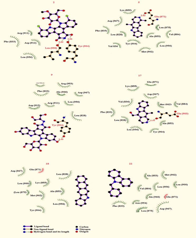
Figure 2. Coupling carbazole derivatives (2, 5, 9, 17, 18, and 22) with the 3pjc protein surface.
Numerous computational methods are available for forecasting the pharmacokinetic behavior of compounds with anticancer potential. For instance, one study explored the pharmacokinetic characteristics of various carbazole derivatives using theoretical tools to assess their likelihood of exhibiting oral bioactivity in humans [44]. In another case, compound SR13668 was evaluated for its pharmacokinetic profile using data from software version 10.2 [45]. These findings highlight how in silico platforms can serve as effective predictors of absorption, distribution, metabolism, and excretion (ADME) properties. Accordingly, the present research focused on examining the pharmacokinetic traits of carbazole analogs 2, 5, 9, 17, 18, and 22 through the SwissADME software, as detailed in Table 3. The analysis indicated that interactions with various cytochrome P450 enzymes may influence metabolic differences among these analogs. Notably, compound 2 exhibited comparatively poor gastrointestinal absorption relative to the other analogs assessed. In addition, this compound demonstrated the potential to inhibit CYP2C9, an enzyme involved in the metabolism of several clinically important drugs. Such inhibition could alter the pharmacological effects of CYP2C9 substrates, including nonsteroidal anti-inflammatory drugs such as celecoxib and flurbiprofen [46], as well as antidiabetic agents such as glibenclamide and gliclazide [47].
Table 3. Pharmacokinetic factors for carbazole analogs 2, 5, 9, 17, 18, and 22
Parameter | 2 | 5 | 9 | 17 | 18 | 22 |
GI absorption BBB permeant P-GP substrate CYP1A2 inhibitor CYP2C19 inhibitor CYP2C9 inhibitor CYP2D6 inhibitor CYP3A4 inhibitor Consensus LogPO/W | Low No No No No Yes No No 2.75 | High Yes Yes Yes Yes No Yes Yes 3.84 | Low No No No No No No No -0.77 | High Yes Yes Yes Yes No Yes Yes 3.14 | High No Yes Yes No No No No 4.82 | High Yes Yes Yes Yes No Yes Yes 4.48 |
Prior research has indicated that certain carbazole analogs may exert toxic effects in various biological systems [48, 49]. For instance, one study reported that a carbazol-oxadiazol compound displayed significantly greater toxicity than its carbazol-acetyl counterpart [42]. Building on these insights, the present study employed the Gussar program to assess the toxicological profiles of carbazole analogs 2, 5, 9, 17, 18, and 22. Findings revealed that compound 9 necessitated a comparatively higher intraperitoneal dose to elicit toxic effects, unlike the other analogs examined. This variation underscores the influence of both dosage levels and drug-delivery methods on the overall toxicity profile of these compounds (Table 4).
Table 4. Toxicity degree for carbazole analogs 2, 5, 9, 17, 18, and 20
Compound | IP LD50 (mg/kg) | IV LD50 (mg/kg) | Oral LD50 (mg/kg) | SC LD50 (mg/kg) |
2 | 552.70 | 558.60 | 5621.00 | 3130.00 |
5 | 429.10 | 67.01 | 649.30 | 190.20 |
9 | 871.00 | 719.20 | 3459.00 | 2179.00 |
17 | 800.10 | 76.74 | 1278.00 | 208.70 |
18 | 305.20 | 72.81 | 1328.00 | 217.60 |
22 | 355.80 | 45.41 | 2194.00 | 723.50 |
IP = intraperitoneal route of administration; IV = intravenous route of administration; Oral = oral route of administration; SC = subcutaneous route of administration
This study explored the theoretical interaction between carbazole analogs and the JAK3 receptor, utilizing the 3pjc protein model as a molecular docking framework. Among the tested compounds, analogs 2, 5, 9, 17, 18, and 22 demonstrated a notably stronger binding potential to the JAK3 surface compared to analogs 1, 3, 4–6, 6–8, 10–16, 19, 20, and 23–26. These computational insights suggest that analogs 2, 5, 9, 17, 18, and 22 may function as effective JAK3 antagonists, potentially suppressing cancer cell proliferation and serving as potential candidates for anticancer drug development.
None
None
None
All experimental protocols adhered to the guidelines established by the Pharmacochemistry Laboratory at the University Autonomous of Campeche.
Open Access The author(s) retain copyright. This article is licensed under the Creative Commons Attribution-NonCommercial-ShareAlike 4.0 International License. It may be shared and adapted for non-commercial purposes with appropriate attribution, an indication of changes, and distribution of adaptations under the same license. Third-party material may be subject to separate terms identified in its credit line. View the license at https://creativecommons.org/licenses/by-nc-sa/4.0/.
 Publication System