Assessing erythrocyte acid resistance is a key part of understanding the effects of toxicants on the blood. The objective of this research is to examine both the isolated and combined effects of cadmium, lead, and zinc ions from contaminated drinking water on the acid resistance of erythrocytes in laboratory rats. This investigation was conducted in the Laboratory of Anatomy, Physiology, and Histology at Chechen State University in Grozny, Russia. The study used laboratory rats weighing 100-150 grams, bred in the university’s vivarium. Exposure to metals altered erythrograms, with a noticeable increase in the proportion of erythrocytes with lower resistance and a reduction in hemolysis time. The most considerable alterations were observed after prolonged exposure to Pb2+, Cd2+, Zn2+, and their mixture. After 30 days of exposure to these ions, the peak times for erythrograms were recorded as 0.5 minutes for Pb2+, 1.0 minutes for Zn2+, and 1.5 minutes for Cd2+. The percentage of erythrocytes undergoing hemolysis at these times was significantly higher, being three times more than the control for Pb2+ and Zn2+, and comparable to the control for Cd2+ (36%). Hemolysis times were notably shorter—2.5 minutes for Pb2+ and Zn2+, and 4.5 minutes for Cd2+. By the end of the 30 days, all rats in the heavy-metal exposure group had died. The findings indicate that prolonged exposure to heavy metals induces significant changes in the erythrocyte population and their acid resistance.
The blood system exhibits both quantitative and qualitative changes in response to various external and internal factors to maintain homeostasis [1, 2]. Toxic exposures can lead to decreased blood and tissue oxygen levels, which, in turn, cause morphological and functional disturbances in erythrocyte membranes [3, 4]. These toxic agents promote the production of free radicals, which leads to oxidative stress and subsequent damage to cell membranes [5-7].
Erythrocytes are an essential indicator of cell membrane integrity, and their functional role is vital for assessing cellular health [8-10]. Understanding the effects of toxic exposure on the blood system helps uncover the compensatory and adaptive mechanisms that occur during chronic exposure to heavy metals. This is valuable not only for analyzing the erythrocyte population during acute toxicity but also for monitoring long-term compensatory processes within the blood system [11-15]. For example, cadmium (Cd2+) can trigger lipid peroxidation and replace iron in heme proteins, while lead (Pb2+) binds to protein and non-protein thiols, disrupting cellular functions [16, 17]. Hosseini et al. [18] showed that tryptophan administration before zinc sulfate exposure reduced erythrocyte hemolysis, but no such effect was noted when lead sulfate was introduced, indicating that Pb2+ or Cd2+ ions might interact more strongly with erythrocyte membrane proteins [19, 20].
Given these observations, assessing erythrocyte acid resistance is a key part of understanding the effects of toxicants on the blood. The objective of this research is to examine both the isolated and combined effects of cadmium, lead, and zinc ions from contaminated drinking water on the acid resistance of erythrocytes in laboratory rats.
This investigation was conducted in the Laboratory of Anatomy, Physiology, and Histology at Chechen State University in Grozny, Russia. The study used laboratory rats weighing 100-150 grams, bred in the university’s vivarium.
The following heavy metals were introduced to the rats in chronic exposure experiments:
1. Group 1: Cadmium chloride at a concentration of 0.25 mg/L (with an established maximum permissible concentration of 0.005 mg/L) [21];
2. Group 2: Lead acetate at a concentration of 0.5 mg/L (maximum permissible concentration being 0.1 mg/L) [22];
3. Group 3: Zinc sulfate at a concentration of 0.1 mg/L (with the permissible limit of 0.01 mg/L) [23];
4. Group 4: A combination of 1.0 milligrams per liter lead acetate, 0.05 milligrams per liter cadmium chloride, and 0.1 milligrams per liter zinc sulfate.
Rats housed in standard vivarium conditions served as the control group. Each experimental group was composed of 20 rats. The acid resistance of erythrocytes was analyzed in the peripheral blood samples of the rats at intervals of five, fifteen, thirty, and forty days, during which they were exposed to water contaminated with the three heavy metals [24]. The results of the study were statistically analyzed using STATISTICA 12.0.
Figures 1-4 display the findings of this study. The analysis reveals that exposure to heavy metal ions, whether in isolation or in combination, induces significant alterations in the structure and function of rat erythrocyte membranes.
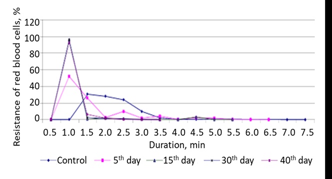
Figure 1. Variation in the acid resistance of rat erythrocytes as a function of the duration of exposure to drinking water contaminated with lead acetate.
The acid erythrograms in the experimental groups were compared based on hemolysis duration, peak timing, and erythrogram characteristics [25]. A leftward shift in the erythrogram was observed on the fifth day of Pb2+ and Cd2+ exposure (Figures 1 and 3), while a mixture of Pb2+, Cd2+, and Zn2+ ions resulted in a shift of 0.5 minutes (Figure 4). As the exposure period extended, more erythrocytes underwent hemolysis at the peak of the erythrogram: 51.9% for Pb2+, 57.1% for Zn2+ (Figure 2), 36.0% for Cd2+, and 76.0% for the combined metal ions. The hemolysis time also changed: with Cd2+ exposure, it lasted 4.5 minutes, whereas the groups exposed to Pb2+ and the metal mixture had the smallest reduction, with hemolysis persisting for 5.5 minutes.
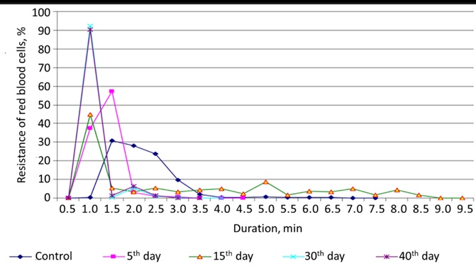
Figure 2. The resistance of rat red blood cells to zinc sulfate changes according to the duration of intoxication through drinking water.
On the fifteenth day of exposure to heavy metal salts, a leftward shift in the erythrogram was noted. Under the influence of Zn2+ ions, the erythrogram also shifted to the first minute, while Cd2+ ions led to a rightward shift by two minutes. Additionally, there was an observed rise in the number of erythrocytes undergoing hemolysis at the peak of the erythrogram.
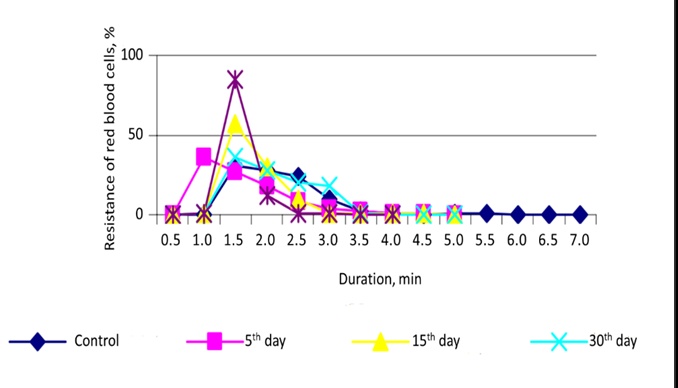
Figure 3. Variation in the resistance of rat erythrocytes to cadmium chloride correlates with the length of drinking water intoxication.
At this phase of metal exposure, nearly 96.0% of erythrocytes underwent lysis following treatment with Pb2+ ions or a combination of multiple metals. When rats were subjected to Zn2+ and Cd2+ ions individually, hemolysis affected 45.0% and 55.0% of the erythrocyte population, respectively. The hemolytic process occurred with marked rapidity—reaching completion in only 1.5 minutes—when induced by the mixed-metal solution. In contrast, Zn2+ exposure prolonged the onset of hemolysis, with the process extending up to 8.5 minutes. Treatment with lead and cadmium salts, on the other hand, resulted in shorter hemolysis intervals of 4.5 and 4.0 minutes, respectively.
Following 30 days of continuous intoxication with Pb2+, Zn2+, and Cd2+ ions, the erythrogram peaks were observed at 0.5, 1.0, and 1.5 minutes, in that order. The extent of hemolysis at these peak points was strikingly elevated—around threefold higher than control levels—for the groups exposed to Pb2+ and Zn2+ ions. Interestingly, the percentage of erythrocytes lysed in the Cd2+ group remained relatively consistent with the control at 36.0%. Hemolysis durations were also considerably reduced under prolonged metal exposure, measuring 2.5 minutes in the Pb2+ and Zn2+ groups and 4.5 minutes in the Cd2+ group. By the end of the 30-day study, all animals consuming water contaminated with heavy metals had succumbed.
Collectively, the data highlight pronounced disruptions in the erythrocyte population of rats exposed to prolonged heavy metal salts. A dominant presence of erythrocytes exhibiting reduced acid resistance reflects advanced cellular senescence, likely stemming from degenerative alterations in membrane structure under chronic intoxication by cadmium, lead, zinc, or their combinations [26–28]. This accelerated erythrocyte aging likely reflects underlying pathological mechanisms, including suppression of erythropoiesis and compromised membrane integrity and permeability, potentially linked to increased lipid peroxidation and diminished antioxidant enzyme function [29, 30].
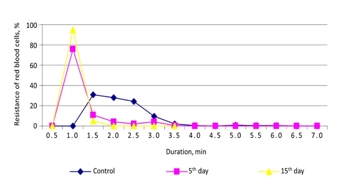
Figure 4. Alterations in the stability of rat blood erythrocytes under the combined impact of heavy metals—cadmium, lead, and manganese—in relation to the length of aquatic environment intoxication.
The present work highlights how examining the progression of acid-induced hemolysis during sustained exposure to heavy metal salts can serve as a reliable indicator of both erythrocyte membrane compromise and disturbances in hematopoietic organ performance. After thirty days of continuous intoxication with Pb2+, Zn2+, and Cd2+ ions, the erythrogram peaks were markedly displaced, occurring at 0.5, 1.0, and 1.5 minutes, respectively. A substantial increase in the proportion of hemolyzed erythrocytes was observed in the Pb2+ and Zn2+ groups, with values reaching nearly threefold those seen in the control group. Conversely, rats exposed to Cd2+ ions displayed hemolytic responses similar to baseline (36.0%). The timeline for hemolytic onset shortened significantly: down to 2.5 minutes for both Pb2+ and Zn2+, and 4.5 minutes for Cd2+. Alarmingly, all animals exposed to metal-contaminated drinking water succumbed by day 30. These findings point to a dramatic alteration in red blood cell dynamics under chronic heavy metal influence, suggesting accelerated senescence and compromised membrane integrity as a consequence of long-term toxic exposure.
None
None
None
All experimental protocols adhered to the European Convention on the Protection of Vertebrate Animals used for Experimental and other Scientific Purposes.
Open Access The author(s) retain copyright. This article is licensed under the Creative Commons Attribution-NonCommercial-ShareAlike 4.0 International License. It may be shared and adapted for non-commercial purposes with appropriate attribution, an indication of changes, and distribution of adaptations under the same license. Third-party material may be subject to separate terms identified in its credit line. View the license at https://creativecommons.org/licenses/by-nc-sa/4.0/.
 Publication System