Parkinson’s disease stands as the second most common neurodegenerative disorder worldwide. This study aimed to assess the impact of DMSO in a rotenone-induced rat model of Parkinson’s disease. DMSO has become a popular agent in preclinical and clinical studies due to its ability to facilitate the transport of poorly soluble drugs across the blood-brain barrier. In this investigation, we explored how a three-week treatment with rotenone, combined with DMSO, influenced hippocampal neuronal activity and the properties of neuronal responses in rats. We specifically compared the toxic effects of rotenone on hippocampal CA1 and CA3 neurons in the presence of DMSO. Our results showed that rotenone induced substantial morphological changes in hippocampal cells. Following DMSO treatment, however, there was a significant restoration of pyramidal cells and Nissl bodies within the CA1 and CA3 regions. DMSO also effectively suppressed both outward and inward currents. Additionally, we recorded spontaneous and evoked spike activity in the hippocampus of rats treated with DMSO (1 ml/kg, administered intraperitoneally for 3 weeks). While rotenone elevated TP and produced a moderate TD effect, DMSO also increased TP but produced a more pronounced TD effect. The analysis indicated inhibitory responses in the hippocampus following high-frequency stimulation (100 Hz for 1 second) of the ipsilateral entorhinal cortex.
Parkinson’s disease (PD) ranks as the 2nd most common neurodegenerative condition associated with aging. The rotenone rat model of PD is a widely used research tool that has been extensively studied for its ability to simulate key aspects of the disease [1]. In this model, rotenone induces motor deficits, though it does not cause significant damage to dopaminergic neurons [2, 3]. Research has identified the hippocampus as a critical region for the memory impairments observed in Parkinson’s disease, with both functional and structural abnormalities in this area reported in patients with sporadic [4, 5] and inherited forms of the disease [4, 6]. Furthermore, dysfunction in the hippocampus is strongly associated with cognitive decline and behavioral changes, including memory deficits [7, 8]. Dimethyl sulfoxide (DMSO) is frequently employed as a solvent to deliver drugs that are poorly soluble in water. It is commonly used in both in vivo and in vitro neuroscience studies due to its ability to dissolve various pharmacological compounds [9]. Numerous studies have highlighted the biological impacts of DMSO, particularly within neurology [10]. For example, DMSO inhibits key ion currents such as Na+, K+, and Ca2+ [11], and it has been shown to alter membrane permeability by blocking the effects of neurotransmitters like acetylcholine, glutamate, and GABA in Aplysia ganglion cells [12]. DMSO also reduces NMDA receptor activation and suppresses both NMDA and AMPA currents in hippocampal neurons [9]. Known for its ability to cross the blood-brain barrier, DMSO has therapeutic potential in conditions such as traumatic brain injury, where it lowers intracranial pressure and improves cerebral blood flow without affecting blood pressure [13]. This study aims to evaluate hippocampal neuronal activity in a rotenone-induced Parkinson’s disease model and to investigate the effects of DMSO treatment on male rats following rotenone exposure.
A total of ten male Wistar rats, each weighing between 200 and 240 grams, were used in this experiment. They were housed in polycarbonate cages with 5 rats per cage in a climate-controlled room maintained at 24 °C, 45% humidity, and a 12-hour light/dark cycle. The animals’ body weight was monitored daily throughout the study.
The research was conducted in accordance with the European Communities Council Directive (2010/63/UE) and received approval from the Ethics Committee of Yerevan State Medical University (Approval code: N4 IRB APPROVAL, November 15, 2018).
The rats were randomly divided into two groups. Group A received rotenone (2.5 mg/kg) subcutaneously every other day for 21 days, with sunflower oil (Sigma-Aldrich) as the solvent. In contrast, group B received 1% DMSO (Sigma-Aldrich, St. Louis, MO, USA; 1 milliliter per kilogram; i.p.) for 21 days (3 weeks of rotenone administration followed by 3 weeks of DMSO treatment).
At the conclusion of the 6 weeks, the rats were euthanized using a deep urethane anesthesia (1.1 grams per kilogram, i.p.). To immobilize the animals, 1% dithylinum (25 mg/kg, i.p.) was used. After anesthesia, the rats were positioned in a stereotactic apparatus and maintained on artificial respiration. A 3 M KCl-filled microelectrode was stereotactically inserted into the hippocampus to record extracellular neuronal activity, following brain atlas coordinates (AP – 3.2-3.5; L ± 1.5–3.5; DV +2.8–4.0 mm) [14]. High-frequency stimulation (HFS, one hundred Hz for one second) was delivered to the contralateral entorhinal cortex using bipolar silver electrodes, with rectangular pulses of 0.05 ms duration and 0.6–0.8 mA amplitude (Figure 1). The coordinates for the stimulation electrode in the ipsilateral entorhinal cortex were set to AP -9, L 3.5, and DV +4.0 millimeters. Data analysis of interspike intervals and spike frequencies was performed using the Student’s t-test and Mann-Whitney U test, as previously outlined [15].
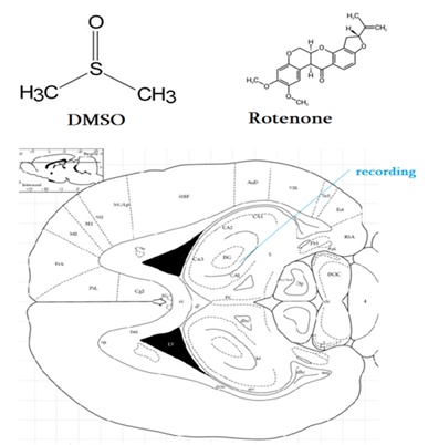
Figure 1. Depictions of Rotenone and DMSO molecular structures. The rat hippocampus is shown using the stereotaxic coordinates from Watson and Paxinos. A blue line marks the electrode positioning.
Following the rotenone and DMSO treatments, the rats were euthanized six weeks later, and their brains were preserved in a 4% formalin solution in phosphate buffer (pH = 7.4) after each electrophysiological assessment. The hippocampus was sliced into serial frozen sections, which were first washed twice with phosphate buffer, then immersed in a 0.5% cresyl violet acid solution for 30 minutes. The sections were then processed through ethanol solutions of 70%, 95%, and 100%, followed by a rinse in distilled water. Afterward, the slices were cleaned twice in xylene, each for five minutes. The tissue was then mounted with an organic medium composed of polystyrene, dibutyl phthalate, and xylene. Histological images of the hippocampus were captured using a light microscope and digital camera after the slices had dried. The rat brain atlas was used to examine histological sections [16, 17].
The detailed characteristics of these cells have been described earlier [15]. To investigate the effects of rotenone and dimethyl sulfoxide (DMSO), extracellular recordings were performed on hippocampal neurons. In the rotenone-treated group, tetanic potentiation (TP) in the hippocampus was observed to be 2.65 times higher (MTT = 39.69 / MBE = 14.96 spikes per second) during high-frequency stimulation (one hundred Hz for one second) as compared to the baseline (Figure 2a), while the TP PTP responses were 1.96 times greater (MTT = 23.39 / MBE = 11.93 spike/sec). There was a significant difference in TP responses between the rotenone and DMSO groups (P < 0.05). For tetanic depression (TD) during HFS, the rate of occurrence was four times higher in neurons showing TD PTP responses (MBE = 35.69 / MTT = 8.91 spikes/sec) and five times higher in neurons exhibiting TD responses (MBE = 42.18 / MTT = 8.42 spikes per second) in the DMSO group (Figure 2b).
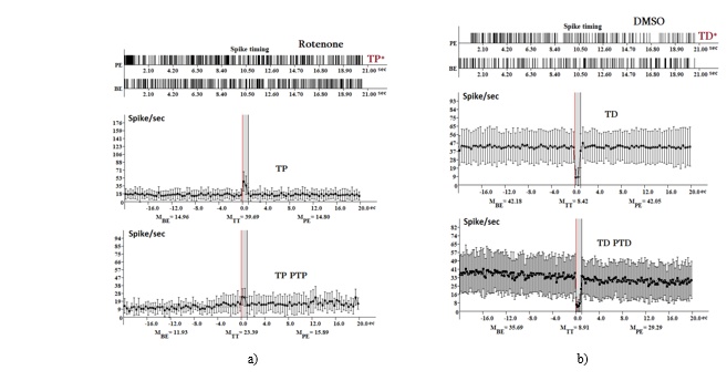
Figure 2. Influence of rotenone and DMSO on neuronal firing in the hippocampus. The spike activity of a single neuron is displayed for 20 seconds before (BE) and after (PE) high-frequency stimulation (HFS), highlighting TP* and TD* responses. Neurons exhibiting TP, TP PTP, TD, and TD PTD responses were analyzed for their average spike rate at three different time points: 20 seconds before stimulation (M BE), during stimulation (M HFS), and 20 seconds after stimulation (M PE).
Rotenone treatment induced marked morphological variations in the hippocampal neurons. Although the neurons retained their staining properties following rotenone exposure, they became noticeably wrinkled. Irregular staining patterns were evident in the hippocampal cells [18]. The Nissl substance in neurons was disrupted, and some cells showed edema. A shift in the distribution of tigroids to the outer edges of specific neurons was observed. Most of the neurons in the rotenone group showed signs of pyknosis (Figure 3). These pyknotic neurons were characterized by their reduced size, distinct oblong shape, and accumulation of dense chromatophilic material (or Nissl bodies) in the cytoplasm. Their nuclei appeared diffusely basophilic, elongated, or triangular, with nucleoli barely detectable.
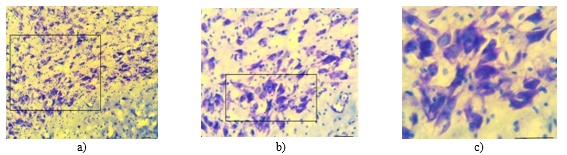
Figure 3. Nissl staining of hippocampal neurons from the rotenone group. Scale bar: A-C 100 µm.
As depicted in Figure 4, neurons in the DMSO group exhibited round to oval-shaped nuclei. The chromatophilic substances within these neurons were distributed throughout the cytoplasm in small clumps with lighter spaces between them. The hippocampal morphology in the DMSO group was characterized by both hyperchromic and hypochromic cells. Neuronal processes extended over short distances, with cell outlines clearly visible, and some neurons displayed light-colored nuclei. Overall, the condition of the nervous tissue showed varying levels of functional activity, indicated by the presence of hyperchromic neurons (Figure 4).
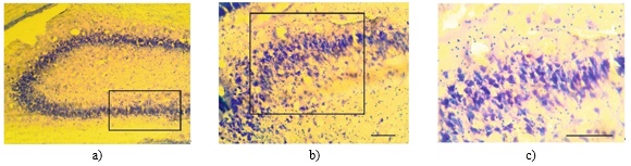
Figure 4. Nissl staining of hippocampal neurons from rats treated with DMSO. Scale bar: A-C 100 µm.
This study explored the effects of 3 weeks of rotenone and DMSO treatment on hippocampal neuronal activity and response patterns in rats. We compared the impact of rotenone on hippocampal CA1 and CA3 cells pre-treated with DMSO. In the DMSO-treated group, hippocampal neurons appeared well-formed, with clearly visible nucleoli, lightly colored cytoplasm, and an organized structure. In contrast, the rotenone-treated group displayed significant cellular damage, including shrinkage, irregular cell shapes, membrane contraction, darkly stained pyknotic nuclei, dispersed neurons, and fewer pyramidal cells and Nissl bodies. However, following DMSO treatment, the pyramidal cells and Nissl bodies in the CA1 and CA3 regions of the hippocampus in the rotenone group displayed substantial recovery (Figures 3 and 4).
From these observations, it can be concluded that rotenone, which inhibits mitochondrial complex I, exerts a different influence on hippocampal synaptic plasticity compared to DMSO. The rotenone group exhibited greater tetanic potentiation, suggesting increased synaptic strength and plasticity following high-frequency stimulation, implying that rotenone might enhance the excitability of hippocampal neurons. Conversely, the DMSO group showed stronger inhibitory responses to high-frequency stimulation (HFS) than the rotenone group (Figure 2), whereas their excitatory responses to HFS were less pronounced. TD and TD PTD were significantly present in the hippocampal neurons (65% and 30%, respectively). These results indicate that DMSO treatment enhanced inhibitory signaling, thereby counterbalancing the heightened neuronal excitability induced by rotenone. The disruption in synaptic balance between excitation and inhibition enabled the system to adjust to new pathological challenges. The dynamic nature of excitation/inhibition allows the neural circuit to operate in a switch-like fashion, amplifying brief high-frequency bursts and transmitting signals with place-field-like characteristics [19]. Excitatory input supports spike integration, while feed-forward inhibition prevents it, reflecting two opposing plasticity mechanisms [20]. In prior experiments using the same rotenone-induced Parkinson’s disease model, we observed that hippocampal neurons favored excitatory activity during high-frequency stimulation of the entorhinal cortex [21].
DMSO is a solvent often used for its ability to mix with water and enhance the permeability of biological membranes, facilitating the absorption of substances that would not otherwise dissolve in water [22]. The impact of DMSO varies depending on factors like the type of cells involved, the specific experimental setup, and its concentration. When tested in cell cultures, DMSO has been demonstrated to alter the excitability of cells by influencing the movement of key ions such as Ca+2, Na+, K+, and Cl- [23, 24]. It has been reported to suppress currents mediated by NMDA and AMPA receptors while decreasing NMDA receptor activation in cultured neurons [9]. In contrast, DMSO inhibits GABA-induced currents in dorsal ganglion neurons from rats [24].
Furthermore, studies conducted both in vitro and in vivo have shown that DMSO administration increases dendritic spine density in a region-specific manner in the hippocampus of APPSDL mice. Behavioral effects of DMSO include improvements in hippocampal-dependent spatial memory accuracy, modulation of olfactory habituation independent of the hippocampus, and reductions in anxiety-like behavior [25]. In adult rats, DMSO treatment has been linked to behavioral alterations thought to reflect changes in hippocampal or cortical activity [26, 27].
As a neuroprotective agent, DMSO is particularly beneficial in severe brain injury, where it can help protect against secondary neuronal death [28]. Its use has been associated with a specific increase in hippocampal spine density in APPSDL mice, both in vivo and ex vivo. Drugs that block the excessive influx of Na+ into brain cells have demonstrated notable neuroprotective effects in models of brain ischemia and hypoxia, with ongoing clinical trials examining their potential in treating cerebral ischemia [29, 30]. DMSO’s neuroprotective properties are believed to stem from its ability to block Na+ channels, thereby helping prevent cellular damage in the brain following injury or ischemia [31]. Modulation of D1/D5 receptors has been shown to affect long-term potentiation in the CA1 region, which is essential for memory processes. This suggests that reduced dopamine signaling in the hippocampus could impair LTP, potentially contributing to memory issues seen in Parkinson’s disease [32, 33]. Additionally, rotenone has been observed to reduce the delayed rectifier K+ current [34], while enhancing the ATP-sensitive K+ current [35] and Ca2+-activated K+ channels with high conductance [36]. Furthermore, rotenone increases NMDA-induced currents in dopaminergic neurons of the substantia nigra [37, 38]. The CA1 neurons in the hippocampus and the striatum are highly susceptible to stress caused by ischemia and metabolic disruptions [38, 39].
Surprisingly, inhibiting GABAergic signaling proves detrimental, especially under conditions of energy scarcity, and enhancing GABAergic modulation could be a potential neuroprotective approach in scenarios with high metabolic demands [39]. Co-activation of GABA(A) and GABA(B) receptors has been shown to protect against ischemia-induced neurodegeneration in vitro. Earlier studies suggested that rotenone’s inhibition of mitochondrial complex I impacts the hippocampus less severely than the striatum [40]; however, increasing evidence aligns with our findings that the hippocampus is notably sensitive to rotenone toxicity [21]. As a critical brain region involved in numerous cognitive functions, including learning and memory, the hippocampus is highly vulnerable to oxidative stress. Mitochondrial byproducts, such as ATP and reactive oxygen species (ROS), are pivotal for maintaining hippocampal synaptic transmission [41]. In the mHippoE-18 model, rotenone induces neuronal death in hippocampal cells [42]. Disruptions in hippocampal long-term potentiation (LTP) and impairments in hippocampal-dependent memory were observed in transgenic mice expressing human alpha-synuclein (alpha-syn120) under the tyrosine hydroxylase promoter [43]. Collectively, these results suggest that both rotenone and DMSO influence synaptic plasticity in the hippocampus, though they exert opposite effects on TP and TD responses. While rotenone enhances TP and induces a milder TD response, DMSO boosts TP while eliciting a more pronounced TD response. This suggests that rotenone and DMSO may exert divergent effects on hippocampal neuronal excitability and plasticity [44]. Rotenone amplifies TP and produces a milder TD reaction, while DMSO boosts TP but causes a more pronounced TD effect. This suggests that the effects of rotenone and DMSO on hippocampal neurons’ excitability and synaptic plasticity differ significantly.
To conclude, the rotenone-treated rat model mimicked the functional deficits characteristic of Parkinson’s disease, particularly affecting hippocampal neurons, whereas DMSO administration altered their excitability.
We extend our thanks to Dr. Susanna Hakobyan for her valuable contribution to the histological work.
None
None
The study adhered to the ethical standards outlined in the European Communities Council Directive (2010/63/UE) and was approved by the Ethics Committee of Yerevan State Medical University under approval code N4 IRB APPROVAL on November 15, 2018.
Open Access The author(s) retain copyright. This article is licensed under the Creative Commons Attribution-NonCommercial-ShareAlike 4.0 International License. It may be shared and adapted for non-commercial purposes with appropriate attribution, an indication of changes, and distribution of adaptations under the same license. Third-party material may be subject to separate terms identified in its credit line. View the license at https://creativecommons.org/licenses/by-nc-sa/4.0/.
 Publication System