Testicular toxicity is recognized as an underlying factor contributing to male infertility. This study evaluated the protective effects of Artemisia herba-alba against calcium tetrachloride (CCl₄)-induced toxicity in rats, focusing on its impact on ERCC1 gene expression. 20 male Wistar rats were randomly divided into four groups (n = 5 per group). Group I served as the untreated control. Group II received oral CCl₄ (0.4 ml/200g) every other day for three weeks. Group III was administered Artemisia herba-alba (ART) extract orally at 500 mg/kg body weight every other day for three weeks. Group IV was treated with both ART extract (500 mg/kg b.w.) and CCl₄ (0.4 ml/200g) on alternating days over three weeks. Parameters assessed included body weight, relative kidney weight, serum testosterone, tissue oxidative stress markers, ERCC1 gene expression, and testicular histology. The results revealed that CCl₄ exposure led to reduced body weight, lower tissue glutathione (GSH), decreased serum testosterone, elevated lipid peroxidation, upregulated ERCC1 expression, and disrupted testicular histoarchitecture. Conversely, ART co-treatment mitigated these effects, improving testicular histology, downregulating ERCC1 expression, and partially preserving body weight and testosterone levels. Further research with extended treatment periods is recommended to confirm the therapeutic potential of ART in managing testicular toxicity.
The testes, located outside the male body in the groin area, are vital reproductive organs essential for producing sperm, synthesizing male hormones (androgens), and transferring genetic information to female gametes [1, 2].
In recent years, testicular toxicity has emerged as a key factor contributing to rising male infertility rates, raising significant public health concerns. The testes’ environment is naturally low in oxygen, making them particularly sensitive to oxidative stress driven by excessive reactive oxygen species (ROS) [3]. Multiple external influences — including pharmaceutical agents, environmental pollutants, and workplace exposures — can compromise testicular health, disrupt normal function, and lead to toxicity [4].
Carbon tetrachloride (CCl₄) is a synthetic chlorinated hydrocarbon with a distinct sweet odor, once commonly used in cleaning products, fire extinguishers, and as a chemical precursor for refrigerants. Despite its historical uses, CCl₄ is highly toxic, and excessive exposure can cause severe damage to the liver, kidneys, lungs, and nervous system [5, 6]. In the body, CCl₄ is metabolized mainly in the liver, where it interacts with cytochrome P450 enzymes to generate harmful free radicals. These reactive byproducts trigger lipid peroxidation, damaging cellular membranes and impairing mitochondrial, endoplasmic reticulum, and plasma membrane integrity, ultimately leading to cell injury and organ dysfunction [7–9].
Artemisia herba-alba (desert wormwood) is a perennial shrub from the Artemisia genus, comprising around 400 species. Characterized by its silver-green, hairy, bi-pinnate leaves, the plant is widely distributed across regions such as North Africa, the Middle East, India, Spain, the Himalayan foothills, and the deserts of the Sinai Peninsula [10–12]. Traditionally, A. herba-alba has been used in folk medicine for treating a range of ailments. Scientific studies support its therapeutic potential, highlighting properties such as antidiabetic, antimicrobial, antioxidant, antifungal, antihypertensive, neuroprotective, immunomodulatory, antimalarial, and antispasmodic activities [10]. Its phytochemical composition includes sesquiterpene lactones, compounds valued for their anti-inflammatory, antioxidant, anticancer, antibacterial, and antimalarial effects [11, 13].
The Excision Repair Cross-Complementing 1 (ERCC1) gene is pivotal to the DNA repair process, ensuring cellular resilience against DNA damage [14]. Although ERCC1 activity is vital for cell survival, its overexpression in testicular germ cells has been linked to resistance to chemotherapy agents such as cisplatin [15].
Given these considerations, this study set out to explore whether A. herba-alba can counteract CCl₄-induced sperm toxicity in rats and to examine its regulatory effects on ERCC1 gene expression.
Fresh A. herba-alba leaves were sourced from a traditional herbal market in Jeddah, Saudi Arabia. The leaves were thoroughly dried at room temperature and ground into a fine powder. 10 g of this powder was mixed with 500 mL of distilled water, then filtered. The resulting extract was concentrated to 8.5 mg/ml and stored at 4 °C until needed for the experiments.
Adult male Wistar rats, weighing 150-250 g, were purchased from the King Fahd Medical Research Center (King Abdulaziz University, Jeddah, Saudi Arabia). Upon arrival, the animals were housed under standard laboratory conditions with a controlled 12-hour light/dark cycle, given free access to food and water, and allowed to acclimatize for one week. All experimental procedures were reviewed and approved by the Ethics Committee of the College of Medicine, King Abdulaziz University.
Carbon tetrachloride (CCl₄) was obtained from Sigma-Aldrich (Missouri, USA) and diluted with olive oil in a 1:10 ratio. Other chemicals used in the study were of analytical grade and used as received.
After the acclimation period, the rats were randomly divided into four groups, each containing five animals, and treated as follows over a period of three weeks:
Group 1 (control) received no treatment.
Group 2 (CCl₄) administered oral doses of CCl₄ (0.4 ml per 200 g body weight) in olive oil every other day.
Group 3 (ART) treated orally with A. herba-alba extract at 500 mg/kg body weight every other day.
Group 4 (CCl₄ + ART) co-treated with both CCl₄ (0.4 ml/200 g) and A. herba-alba extract (500 mg/kg), administered on alternate days.
At the end of the treatment period, the animals were fasted overnight and then anesthetized using diethyl ether. Blood samples were collected from the abdominal aorta, and the testes were excised, washed in saline, and weighed. Portions of the testicular tissue were fixed in 10% buffered formalin for histological evaluation, while other sections were stored at −80 °C for RNA analysis. Remaining tissue samples were homogenized in 100 mM phosphate buffer (pH = 7.4) and centrifuged at 14,000 rpm for 30 minutes.
Levels of glutathione (GSH) and malondialdehyde (MDA) were assessed in the supernatant using commercially available assay kits (MyBioSource, California, USA), following the manufacturer’s protocols.
Serum testosterone concentrations were determined using an ELISA kit (Diagnostic System Laboratories Inc., USA), as per the supplier’s instructions.
Total RNA was isolated from testicular tissue using the QIAgen RNeasy Mini Kit (cat #74104). For complementary DNA (cDNA) synthesis, 200 ng of RNA was reverse transcribed using the M-MLV Reverse Transcriptase System (Promega, USA). Quantitative PCR reactions were set up with 3 µl cDNA, 0.5 µl of each primer (500 nM), 1 µl of nuclease-free water, and SYBR Green Master Mix (Applied Biosystems, USA). The relative expression levels of target genes were calculated using the 2^−ΔΔCT method, normalized to GAPDH (Table 1).
Table 1. Primer sequences
Isoforms Isoforms | Primers sequence (5`-3`) Primers sequence (5`-3`) |
CYP1A1 F ERCC1 - left | GGG AGG TTA CTG GTT CTG G 5'-AAG GCG TAT GAG CAG AAG C-3’ |
CYP1A1 R ERCC1 right | ATG AGG CTG TCT GTG ATG TC 5'-TCC AAA TGT AGT GAG GAG GGT-3' |
GAPDH F GAPDH - left | GAT GGT GAA GGT CGG TGT G 5'-GAT GGT GAA GGT CGG TGT G-3' |
GAPDH R GAPDH -right | ATG AAG GGG TCG TTG ATG G 5'-ATG AAG GGG TCG TTG ATG G-3' |
Testicular tissues were fixed in 10% buffered formalin, dehydrated in a graded ethanol series, and embedded in paraffin wax for 24 hours at room temperature. Thin sections were sliced from these blocks and stained using hematoxylin and eosin (H&E) to assess histopathological changes. Prepared slides were examined under a light microscope at 400× magnification to capture detailed images of the tissue architecture.
All experimental data were analyzed using one-way ANOVA. The results are expressed as mean ± SEM. Dunnett’s multiple comparisons test was used to evaluate group differences, with P < 0.05 considered statistically significant.
Consistent with prior toxicological reports, CCl₄ exposure significantly reduced (p < 0.05) final body weight in rats compared with untreated controls (Figure 1a). Meanwhile, rats treated with the ART extract alone showed no significant change in body weight compared with controls. Notably, co-treatment with ART in CCl₄-administered rats produced a 17% improvement in body weight over the CCl₄-only group. Although the CCl₄ group showed a 16% decrease in relative testes weight compared with controls, this difference was not statistically significant. Additionally, relative testes weights remained largely unchanged between the CCl₄-only and CCl₄ + ART groups (Figure 1b).
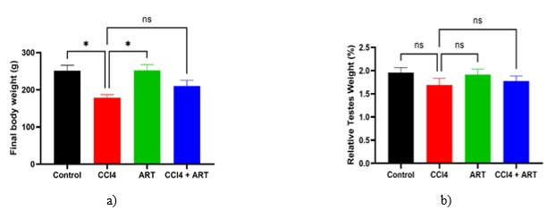
Figure 1. Effects of CCl4 and ART on final body weight and relative testes weight: a) final body weight, and b) relative testes weight.
To evaluate antioxidant status, we measured serum glutathione (GSH) and malondialdehyde (MDA) levels. CCl₄ administration markedly reduced GSH levels (P < 0.01) compared to both the control and ART-only groups (Figure 2a). ART administration alone maintained GSH levels comparable to controls. Interestingly, while ART treatment after CCl₄ exposure led to an 8% increase in GSH relative to the CCl₄-only group, this improvement did not reach statistical significance.
As expected, MDA levels, an indicator of lipid peroxidation, were significantly elevated in the CCl₄ group compared with the control and ART-only groups (Figure 2b). Co-treatment with ART did not considerably reduce MDA levels compared with the CCl₄-only group, suggesting limited reversal of oxidative damage in this marker.
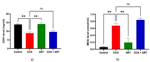
Figure 2. Effects of CCl4 and ART on GSH and MDA levels: a) serum glutathione (GSH) level, and b) serum malondialdehyde (MDA) level.
We next examined the impact on serum testosterone and ERCC1 gene expression in testicular tissue. CCl₄ exposure caused a substantial reduction (P < 0.001) in serum testosterone compared to both the control and ART-only groups (Figure 3a). ART treatment alone preserved testosterone levels comparable to controls, while ART co-treatment in CCl₄-exposed rats led to a 26% increase in testosterone versus CCl₄-only animals — although this rise was not statistically significant.
On the molecular level, ERCC1 gene expression was significantly upregulated in the testes of CCl₄-treated rats compared to controls and the ART-only group (Figure 3b). Notably, ART co-treatment significantly reduced ERCC1 expression (P < 0.0001) in CCl₄-administered rats, bringing levels closer to those in controls.
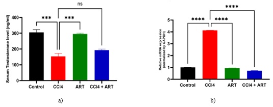
Figure 3. Effects of CCl4 and ART on testosterone levels and ERCC1 gene expression: a) serum testosterone level, and b) relative ERCC1 gene expression.
Histopathological evaluation revealed that CCl₄ exposure disrupted the structural integrity of the testes, as evidenced by disorganized seminiferous tubules and degenerative changes in the testicular membrane, compared to the normal architecture observed in the control group (Figures 4a–4c). The ART-only group displayed healthy, intact seminiferous tubules, similar to those of controls. Remarkably, ART co-treatment after CCl₄ exposure largely restored testicular architecture, with visible improvement in epithelial thickness and increased numbers of primary and secondary spermatocytes (Figures 4d–4e).
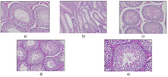
Figure 4. The impact of CCl₄ and ART on testicular histology is shown: (a) testicular sections from the control group display intact seminiferous tubules and normal epithelial lining, (b) testes from the CCl₄-treated group exhibit disrupted membranes and distorted seminiferous tubules, (c) testes from the ART-only group maintain normal histoarchitecture, and (d–e) testes from the group treated with both CCl₄ and ART show near-normal structure, with restored epithelial height and visible spermatogonia.
Previous research has demonstrated that oxidative stress arises when either free radical production exceeds antioxidant defense capacity or antioxidant defense capacity is weakened [16]. For normal cell function, it’s essential to maintain a balance between reactive oxygen species (ROS) and antioxidants [17]. An increase in ROS levels disrupts key cellular antioxidant mechanisms [18]. While mammalian cells contain a variety of enzymatic and non-enzymatic systems to counteract ROS and free radicals, these defenses can sometimes be overwhelmed [19]. As a result, recent attention has focused on antioxidant-rich plants that could help combat ROS-induced oxidative stress [20].
This study evaluated the protective effects of A. herba-alba on CCl₄-induced testicular toxicity. A. herba-alba is known for its antioxidant, antimicrobial, neuroprotective, antimalarial, and immune-modulatory activities [10]. The findings showed that CCl₄ administration led to a reduction in final body weight and a 16% decrease in relative testes weight — findings in line with those of Hashem [17], who observed marked decreases in body and testes weights following CCl₄ exposure due to its toxic effects. Notably, treatment with both CCl₄ and ART resulted in a 17% improvement in body weight (though not statistically significant).
Moreover, the CCl₄-induced reduction in glutathione (GSH) and increase in malondialdehyde (MDA) levels indicate oxidative stress, which can impair spermatogenesis and steroid hormone production [21]. In this study, CCl₄ significantly lowered testicular GSH, consistent with earlier work on CCl₄ toxicity prevention [19]. ART treatment of CCl₄-exposed rats led to a modest (8%) but non-significant rise in GSH, contrasting with the significant boost seen with Launaea procumbens treatment in previous studies [22]. The CCl₄-induced elevation in MDA levels was consistent with Ojo et al. [21], although ART treatment post-CCl₄ did not significantly reduce these elevated levels (Figure 2b), possibly due to the short treatment period.
The significant reduction in serum testosterone levels observed with CCl₄ exposure, as also reported by Shareen et al. [23], suggests either direct Leydig cell damage or indirect disruption via oxidative stress that affects hormonal balance — potentially impairing sperm production. While ART treatment led to a 26% increase in testosterone compared to the CCl₄-only group, this change was not statistically significant.
ERCC1, a key DNA excision repair protein, helps repair chemically induced DNA damage and maintain chromosomal stability [24, 25]. Soares et al. [26] highlighted its association with gastrointestinal toxicity after chemotherapy and radiation in cervical cancer, and other studies have linked ERCC1 status with treatment toxicity and patient-reported outcomes [27]. In this study, CCl₄ significantly upregulated ERCC1 expression in the testes compared with controls and the ART-only group (Figure 3), consistent with Jacobsen et al. [28], who reported that such changes are associated with genomic instability from chemical exposure [26]. ART treatment in CCl₄-administered rats reduced ERCC1 expression.
Histological analysis revealed that CCl₄ disrupted the seminiferous tubules, damaged testis membranes, and reduced spermatogonia. Remarkably, ART treatment after CCl₄ exposure mitigated these structural damages, improving testicular architecture. These findings align with prior studies documenting testicular damage and reduced spermatogenesis following CCl₄ exposure [18, 21, 29].
This study investigated the protective potential of A. herba-alba against CCl₄-induced testicular toxicity in rats. CCl₄ administration led to reductions in body weight, GSH, and testosterone, increased MDA levels, upregulation of ERCC1 expression, and histological damage to the testes. Treatment with A. herba-alba helped restore testicular structure and downregulate ERCC1 expression, while moderately supporting body weight recovery. Further research, particularly with extended treatment durations, is needed to confirm the therapeutic potential of ART for addressing testicular toxicity.
None
None
None
None
Open Access The author(s) retain copyright. This article is licensed under the Creative Commons Attribution-NonCommercial-ShareAlike 4.0 International License. It may be shared and adapted for non-commercial purposes with appropriate attribution, an indication of changes, and distribution of adaptations under the same license. Third-party material may be subject to separate terms identified in its credit line. View the license at https://creativecommons.org/licenses/by-nc-sa/4.0/.
 Publication System