Turmeric (Curcuma longa L.) has antiestrogenic effects that may interfere with the activity of the hypothalamic-pituitary axis, thereby disrupting estrogen production and influencing both uterine weight and diameter. This experiment investigated how turmeric extract affects uterine parameters in female white rats (Rattus norvegicus, Sprague Dawley strain). A total of 28 rats were assigned to four groups: the control group (C) received only H₂O. In contrast, treatment groups T1, T2, and T4 were administered turmeric extract at doses of 250 mg/Kg BW, 500 mg/Kg BW, and 1000 mg/Kg BW, respectively, each combined with 1 ml of H₂O. The treatments were given once daily for five days. Results indicated that turmeric extract at 250 mg/Kg BW and 1000 mg/Kg BW led to a significant reduction in both uterine weight and diameter. In summary, turmeric extract reduced uterine size and weight, indicating its antiestrogenic capability in female rats.
One of the major challenges faced by many developing countries, particularly Indonesia, is the rapid and uncontrolled population growth. Indonesia is ranked fourth among the countries with the largest populations worldwide [1]. To manage population growth, the government has implemented Family Planning programs, which include contraception for married couples [2]. However, the contraceptives currently in use often cause various side effects such as dizziness, fatigue, bleeding, weight gain, hypertension, and menstrual irregularities. Because of this, ongoing research aims to develop contraceptives that are effective, safe, comfortable, reversible, free of side effects, and do not disrupt the menstrual cycle [3].
The use of traditional medicinal plants offers several benefits, including ease of access, affordability, low toxicity, and minimal side effects [4–7]. Among these plants, turmeric (Curcuma longa L.) is considered a potential contraceptive. The primary compound, curcumin, found in turmeric rhizomes, possesses antiestrogenic properties by inhibiting the pituitary gland and hypothalamus, leading to reduced estrogen hormone secretion [8].
Estrogen is a key hormone that regulates the reproductive system, influencing cell proliferation within reproductive organs, such as the uterus. In the uterus, the endometrial layer is highly responsive to fluctuations in estrogen levels. The endometrium consists of epithelial and stromal cells; their proliferation stimulates an increase in cell numbers and enhances the vascularization of spiral arteries, which supply blood to the endometrium, thereby thickening the endometrial layer [9]. Endometrial thickness plays a significant role in determining uterine weight and diameter [10]. Due to its antiestrogenic effects, curcumin can reduce endometrial thickness and subsequently impair implantation capacity [11]. Considering this background, investigating the impact of turmeric extract’s antiestrogenic activity on reducing uterine weight and diameter in female rats is essential. This experiment examined how turmeric extract affects uterine parameters in female white rats (Rattus norvegicus, Sprague Dawley strain).
Turmeric ethanol extract was prepared through the maceration method. Five hundred grams of turmeric powder were soaked in 8 liters of 96% ethanol. The resulting extract was then filtered and concentrated by evaporating with a vacuum rotary evaporator at 50–60 ºC [12].
Twenty-eight female rats aged between 8 and 12 weeks, weighing from 120 to 160 grams, were sourced from the Animal Vet Laboratory Service in Bogor, Indonesia. These rats were randomly assigned to four groups. The control group (C) received only H₂O, whereas treatment groups T1, T2, and T4 were given turmeric rhizome extract doses of 250 mg/kg BW, 500 mg/kg BW, and 1000 mg/kg BW, respectively, each accompanied by 1 ml of H₂O. The extracts were administered orally once daily for five days using a stomach tube. After the treatment period, the stomachs were opened to retrieve the uterus, which was then fixed in 10% buffered formalin. Samples were sent to the Anatomical Pathology Laboratory at the Faculty of Medicine, University of Lampung, for histological examination. The entire study was conducted in accordance with ethical approval from the Health Research Ethics Commission of the Faculty of Medicine, University of Lampung, reference number 3048/UN26.18/PP.05.02.00/2019.
This study focused on uterine histological changes, specifically monitoring uterine weight and diameters in female Rattus norvegicus after treatment with turmeric rhizome extract (C. longa).
SPSS software was used for data analysis, beginning with a bivariate approach. For datasets with 50 samples or fewer, the Shapiro-Wilk test was used to assess normality. If the data met the assumptions of normal distribution and homogeneity, one-way ANOVA was employed for parametric testing. Should differences emerge, post-hoc analysis using the Least Significant Difference (LSD) test was conducted at a 5% significance threshold.
As shown in Table 1, rats receiving turmeric rhizome extract at doses of 250 mg/kg BW and 1000 mg/kg BW exhibited a significant decrease in uterine weight compared to controls (P < 0.024). Similarly, uterine diameters in these groups were significantly reduced relative to the control group (P < 0.000). However, administration of 500 mg/kg BW turmeric rhizome extract resulted in a notable increase in both uterine weight and diameter compared to the control.
Table 1. Effect of turmeric rhizome extract on weight and uterine diameters of rats
Treatments | Parameters (mean ± SD) | |
Uterine weight (g) | Uterine diameters (µm) | |
C (control) | 0.78 ± 0.17ab | 2897.60 ± 296.59ab |
T1 (250 mg) | 0.59 ± 0.23a | 2740.28 ± 364.20c |
T2 (500 mg) | 0.85 ±0.12b | 3031.19 ± 174.37b |
T3 (1000 mg) | 0.67 ±0.04c | 2385.97 ± 113.35ac |
P | 0.024 | 0.000 |
Note: Numbers followed by the same letter indicate no significant difference at the 5% level, the post hoc LSD (least significant difference) test.
The histological images of rats treated with turmeric rhizome extract compared to those untreated are shown in Figure 1.
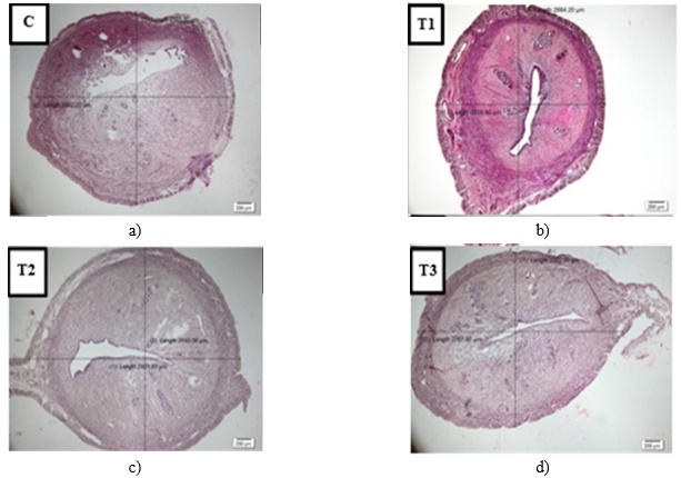
Figure 1. Histological images of the uterus in mice treated and untreated with turmeric extract (C. longa). Description: C = control; T1 = treatment 1; T2 = treatment 2; T3 = treatment 3. Observed under a light microscope at 4 x 10 magnification, stained with Hematoxylin–Eosin.
Based on the findings, administering turmeric extract at doses of 250 mg/kg BW and 1000 mg/kg BW resulted in a decrease in uterine weight. In contrast, a dose of 500 mg/kg BW turmeric rhizome extract caused an increase compared to the control group. Turmeric, known as a traditional medicinal plant, is used as a contraceptive. Its primary active ingredient, curcumin, can suppress the production of estrogen and progesterone. These hormones significantly influence the endometrium, one of the reproductive organs highly sensitive to their levels. Therefore, a reduction in estrogen and progesterone during pregnancy can lead to pregnancy complications. Increased estradiol, a derivative of estrogen, correlates with the thickening of the endometrium. This is supported by Purwanti’s research [13], which demonstrated that curcumin administration reduced endometrial thickness in Rattus norvegicus.
The reduction in uterine thickness is directly proportional to the decrease in uterine weight, mainly because the endometrium—a uterine lining highly responsive to estrogen fluctuations—is affected. Consequently, thinning of the endometrium reduces uterine diameter and lowers uterine weight. This finding aligns with the results of Pratiwi [14], who reported that administering turmeric extract (C. longa) to female white mice resulted in a decrease in their endometrial thickness. A thinner endometrium inhibits implantation, reducing the number of implants and causing infertility in the organism [15].
Estrogen plays a vital role in female reproductive organs through two receptor types: alpha receptors (REα) and beta receptors (REβ). Alpha receptors are located in the uterus, adrenal glands, pituitary, epididymis, testes, and kidneys, while beta receptors are primarily found in the ovaries, bladder, brain, bones, prostate, and lungs [16]. Estrogen exerts its effects by binding to these receptors on target cells, inducing conformational changes that stimulate cellular proliferation and increase endometrial thickness [17]. An increase in uterine weight corresponds with an enlargement in uterine diameter. Additionally, the thickening of the endometrium involves a hypoxic process that elevates mRNA expression coding for VEGF, which promotes angiogenesis, enhancing vascular permeability and vasodilation. Curcumin induces hypoxia by inhibiting COX-2 (Cyclooxygenase-2), suppressing epithelial cell proliferation, promoting apoptosis, and hindering angiogenesis [18].
VEGF is the principal factor driving cyclical angiogenesis in the non-pregnant endometrium, with hypoxia serving as a potent stimulus for new blood vessel formation. Elevated VEGF expression occurs when stromal and glandular cells experience oxygen deprivation, leading to hypoxia and increased transcription of the VEGF gene. This hypoxic response is regulated by HIF1 (hypoxia inducible factor 1), which functions to control the quantity of genes activated as part of the cell’s adaptive reaction to oxygen deficiency [19].
The findings revealed that administering turmeric rhizome extract at 250 mg/kg BW and 1000 mg/kg BW doses significantly reduced the uterine diameters in rats. This effect is mainly due to curcumin. Curcumin contains phytoestrogens that behave like estrogen at low concentrations (estrogenic effect) but inhibit estrogen activity when present at higher levels (antiestrogenic effect). When estrogen levels are elevated, phytoestrogens can bind to estrogen receptors, although their affinity is weaker than that of estrogen itself. This competitive binding blocks estrogen from attaching to its receptors, resulting in antiestrogenic outcomes. Conversely, when estrogen concentrations drop, phytoestrogens dominate receptor binding, exerting estrogen-like effects [20]. This mechanism leads to the observed decrease in uterine diameter.
After puberty, estrogen secretion doubles, regulated by the hypothalamus-pituitary axis. The growth and development of reproductive organs continue with age, with the endometrium being highly responsive to estrogen. Estrogen promotes proliferation of the endometrial stroma, facilitating ovum development during implantation. Progesterone is another hormone that influences the female reproductive organs; it enhances secretory transformations in the uterine lining and prepares the uterus to support a fertilized ovum. Additionally, progesterone reduces both the frequency and strength of uterine contractions, helping to prevent the expulsion of the implanted ovum [21].
Curcumin also possesses antifertility properties by downregulating the expression of the ERβ (Estrogen Receptor β) gene and estrogen production in granulosa cells stimulated by FSH and LH. The reduction of ERβ and estrogen disrupts estrogen’s function, impairing steroidogenesis and folliculogenesis. This disturbance negatively affects cell differentiation, FSH activity, estrogen synthesis, and the expression of LHR (luteinizing hormone receptor) and FSHR (follicle-stimulating hormone receptor) [22].
In conclusion, turmeric rhizome extract (C. longa) administered at doses of 250 mg/kg BW and 1000 mg/kg BW can reduce uterine weight and diameter, demonstrating antiestrogenic potential in female Rattus norvegicus (Sprague-Dawley strain).
None
None
None
None
Open Access The author(s) retain copyright. This article is licensed under the Creative Commons Attribution-NonCommercial-ShareAlike 4.0 International License. It may be shared and adapted for non-commercial purposes with appropriate attribution, an indication of changes, and distribution of adaptations under the same license. Third-party material may be subject to separate terms identified in its credit line. View the license at https://creativecommons.org/licenses/by-nc-sa/4.0/.
 Publication System