The Ebola virus is a highly infectious pathogen with no effective antiviral treatments currently available, prompting ongoing research into potential therapeutic options. This study evaluated the inhibitory effects of licensed non-viral drugs on Ebola virus entry and replication using bioinformatic tools. A descriptive-analytical approach was used, in which the chemical structures of selected drugs were first generated in ChemDraw Ultra 10.0 and then energy-optimized in Hyperchem 8.0. Molecular docking was performed using AutoDock4.2 to simulate interactions between the drugs and viral proteins. The analysis revealed that the interactions involved primarily hydrophobic, π-π stacking, hydrogen bonding, and cation-π interactions. Chloroquine, diphenoxylate, and amodiaquine showed the strongest binding affinity, with the most negative docking energies, indicating their potential as effective inhibitors of the GP and VP40 proteins. Conversely, erythromycin and dirithromycin, due to their high hydrophilicity, exhibited weaker binding results. Overall, the study highlighted that drugs with hydrophobic components, effective hydrogen bonding, and tertiary amines tend to show enhanced anti-Ebola properties. The bioinformatic analysis suggests that these drugs could serve as promising candidates for inhibiting Ebola virus entry and replication.
Ebola virus disease is one of the deadliest viral infections in history, originating in West Africa and rapidly spreading worldwide [1-4]. The virus is primarily transmitted through direct contact with the bodily fluids of infected individuals, leading to immune system suppression, acute hemorrhagic fever, and ultimately death [5, 6]. The Ebola virus was first identified in Congo in 1976, with the virus named after a river in the Democratic Republic of Congo. The initial outbreak resulted in 284 cases and 151 deaths [7-9]. Despite nearly 40 years of knowledge about the virus, its most recent outbreak in West Africa has caused a crisis, with over 28,610 cases and 11,315 deaths by March 2016 [10].
Ebola is classified as a member of the Filoviridae family within the Mononegaviral order. The virus has a lipid envelope, a negative-sense RNA genome, and seven proteins. The filamentous virus has a mature virion diameter of approximately 80 nm and a length of approximately 1200 nm. Ebola is transmitted to humans through animals such as bats, chimpanzees, and gorillas [11-13]. Among the virus’s proteins, glycoprotein (GP) is critical. It is the sole structural glycoprotein, containing 676 amino acids, and undergoes cleavage by the host cell’s furin enzyme to form two subunits, GP1 and GP2, linked by a disulfide bond. GP is located on the virus’s surface and is essential for its attachment to and entry into host cells, making it a key player in the virus’s life cycle [11, 14].
Recent research has increasingly focused on inhibiting GP to prevent the Ebola virus from entering and attaching to host cells [14, 15]. Another key protein is the matrix protein VP40, which is critical for the virus’s structural integrity, maturation, and replication. VP40, containing 326 amino acids and weighing 35 kDa, plays vital roles in virion formation, regulation of viral transcription, and the assembly and budding of new virions. Like GP, VP40 is considered a promising target for anti-Ebola drugs, as inhibitors can interfere with virus replication and assembly [16-18]. Currently, no effective treatment exists for Ebola, which continues to pose a major global health threat [7, 9]. Although antibody therapies have been tested in animal models and administered to a few patients, their availability remains limited [19].
In recent years, drug repositioning—finding new uses for existing licensed drugs—has gained considerable attention in the pharmaceutical industry as an alternative to traditional drug discovery methods. This approach offers several advantages: since the drugs have already passed numerous toxicity and safety tests, the risk of adverse effects is lower, and the high costs associated with new drug development are reduced. In addition, the computational method of structure-based drug design, in which small molecules are “docked” into target macromolecular structures and their binding scores are assessed, is widely employed in drug discovery and optimization. The exploration of Ebola virus inhibitors to reduce patient mortality and improve recovery has been an emerging focus of research in recent years. However, challenges remain in understanding the mechanisms by which inhibitors interact with the Ebola virus, as well as the relationship between their chemical structure and anti-Ebola activity. The drug repositioning strategy has been increasingly used to identify new Ebola inhibitors, with several drugs previously tested for Ebola showing potential [20, 21]. Computational methods, particularly in drug design, have become increasingly popular due to advances in computing power, enabling researchers to predict biological activities, discover new compounds, and better understand molecular interactions.
Additionally, these computational methods enable the prediction and analysis of how a compound interacts with a target protein, the evaluation of energy differences among the compound’s various conformations, and the examination of reaction mechanisms. These methods also help in understanding the role of different groups and substitutions within the chemical structure of the compounds and their impact on binding efficiency. However, molecular docking simulations have not yet been applied to licensed drugs that have demonstrated anti-Ebola activity, particularly regarding how these compounds interact with the Ebola virus glycoprotein (GP) and matrix protein (VP40). To address this gap, the current research investigates the drug-receptor interaction energy and the involvement of specific amino acids in the binding process. Furthermore, the study explores the relationship between structural modifications in the drugs and their performance in inhibiting GP and VP40 proteins.
Molecular docking was performed using AutoDock4.2 software. For this purpose, AutoCAD was installed on an 8-core Windows computer to run the simulations. In this study, the interactions of 24 FDA-approved drugs, previously identified as having anti-Ebola effects through various experimental studies [20], with the amino acids of the GP and VP40 binding sites were analyzed using molecular docking simulations. The drug names and their respective IC50 values are listed in Table 1.
Table 1. Drug name and IC50 values against the Ebola virus.
No. | Medicine name | Concentration (micromolar) | Assessment of viral replication | Evaluation of virus-like entry (% activity) |
1 | Amlodipine | 10 | - | 93 |
2 | Amodiaquine | 10 | - | 99 |
3 | Biperiden | 50 | - | 98 |
4 | Carprofen | 10 | - | 94 |
5 | Chloroquinone | 50 | + | 97 |
6 | Dex brom pheniramine | 50 | - | 85 |
7 | D Bucain | 10 | - | 99 |
8 | Diphenoxylate | 50 | + | 96 |
9 | Diphenylpyraline | 50 | + | 96 |
10 | Dirithromycin | 50 | - | 99 |
11 | Erythromycin | 10 | - | 96 |
12 | Estradiol | 10 | - | 93 |
13 | Fluoxetine | 10 | - | 96 |
14 | Ketotifen | 50 | + | 99 |
15 | Levopropoxyphene | 50 | - | 98 |
16 | Mycophenolate | 50 | - | 91 |
17 | Oxyphencyclamine | 50 | - | 95 |
18 | Paroxetine | 10 | - | 98 |
19 | Penbutolol | 10 | - | 98 |
20 | Prochlorperazine | 10 | - | 95 |
21 | Protriptyline | 10 | - | 83 |
22 | Toremifene | 10 | - | 97 |
23 | Dipivefrine | 50 | - | 94 |
24 | Trihexyphenidyl | 10 | - | 97 |
Molecular docking simulations were performed using AutoDock4.2, with the genetic algorithm (GA) used for the search. The docking simulations were prepared, executed, and analyzed using AutoDock Tools (ADT) 1.5.6. To begin, the chemical structures of the drugs were drawn in ChemDraw Ultra 10.0, followed by energy optimization in Hyperchem 8 using the molecular mechanics (MM) force field and the PM3 semi-empirical method. Hydrogen atoms were then incorporated into the ligand structures using AutoDock Tools. In subsequent steps, non-polar hydrogen atoms were merged with their corresponding carbon atoms, and the molecular charges were adjusted to match experimental values. The torsional degrees of freedom for each ligand were also calculated, and the ligand files were saved in pdbqt format.
The 3D crystal structures of the Ebola virus GP and VP40 proteins were retrieved from the protein database. The docking and conformer analysis methods were performed based on previously established protocols [22, 23]. Water molecules were removed from the crystallographic structures using Notcpat++ or Discovery Studio Viewer Lite 4.0, and hydrogen atoms were added with AutoDock Tools. Non-polar hydrogen atoms were integrated with carbon atoms, while the protein’s electric charges and solvent coverage parameters were calculated. The macromolecule files were saved in pdbat format.
Once the required input files for docking (ligand and receptor) were prepared, docking studies were performed to model interactions between the ligand and receptor, using the Lamarckian genetic algorithm. For each ligand, a grid of 60x60x60 Å was generated based on the ligand’s molecular weight, with grid points spaced 0.573 Å apart. The grid centered on the receptor’s active site was saved as a gpf file for AutoGrid calculations. After completing the docking process, the interactions between the ligand and receptor were analyzed, focusing on hydrogen bonds, hydrophobic interactions, and π-π stacking with the amino acids in the receptor’s binding pocket. These analyses were performed using AutoDock Tools and Discovery Studio Viewer Lite 4.0.
Due to the lack of crystallized ligands available for the protein, docking validation was performed through blind docking. In this method, the entire protein was placed in the grid box, and the ligand’s placement and interactions within the protein’s active site were compared with the reported results from earlier studies. Docking of the most effective drug, amodiaquine, was performed, and the ligand was successfully positioned at the active site, exhibiting expected interactions as reported in previous research. Multiple docking attempts showed that the ligand consistently bound to the same site, indicating the active binding region. Once the docking protocol was validated, the analysis of the docking results revealed that all compounds interacted similarly with the receptor’s binding site. The docking data are presented in Table 2.
Table 2. Binding free energy of hydrogen bonds, hydrophobic, π-π, and cation-π interactions of drugs in VP40 protein based on molecular docking.
Medicine name | Binding free energy (kilocalories per mole) | Hydrogen bonds | Hydrophobic interactions | π-π interaction |
Amlodipine | -6.57 | His516, Asn512, Glu106, Trp104 | Glu103, Trp104, Ala105, His518, Arg136, Asn514, Asn512, Trp291, Phe290, Leu547, His516, Glu106 | - |
Amodiaquine | -3.35 | Ser316, Leu158 | Glu103, Trp104, Tyr213, Tyr214, Arg136, Cys135, His516, Ala105, Phe290, Asn514, Asn512 | Tyr214 |
Biperiden | -5.87 | 214Arg | Phe161, Leu158, Phe157, Glu155, Arg148, Ala156, Ser316, Lys212, Cys314, Ser319, Leu213, His315, Pro290 | - |
Carprofen | -7.45 | Cys314, Gln155, 214Arg | Ser319, Ser316, Leu158, His315, Leu213, Lys212 | - |
The evaluation of the best-docked state in molecular docking simulations relies on two essential criteria: the most negative estimated free binding energy and the most favorable interactions with key amino acids in the GP and VP40 receptor active sites. This study performed molecular docking simulations for twenty-four licensed drugs to examine their interactions with GP and VP40. Binding energy and docking energy were quantitatively calculated, incorporating intramolecular energy, torsional free energy, and ligand internal energy [24-26].
The most negative ΔGbinding (binding free energy), as well as favorable interactions and tight binding to key amino acids in the GP active site, were observed for several compounds. Among the analyzed drugs, amodiaquine, diphenoxylate, ketotifen, and paroxetine showed the best docking results, with binding energies of -8.37, -9.13, -8.50, and -8.18 kcal/mol, respectively. A 2013 study by Madrid et al. [20] showed that amodiaquine was the most potent anti-Ebola drug, with 99% inhibition of Ebola virus entry into host cells, supporting the in silico findings of this study. Conversely, Azithromycin and Erythromycin exhibited the lowest binding energies of -4.36 and -5.89 kcal/mol, respectively.
For VP40, the drugs ketotifen and chloroquine demonstrated the highest binding energies. Amodiaquine, which contains a quinoline ring, prevents Ebola virus entry into host cells at a concentration of 10 micromolar, with 99% efficacy. Docking simulations revealed that amodiaquine binds to the GP protein through hydrogen bonding and hydrophobic interactions. The bulky part attached to carbon number 4 of the quinoline ring and the tertiary amine at the end of this group interact hydrophobically with several amino acids, including Asn512, Asn514, His516, Glu71, Glu103, Phe290, and Glu106. The chlorine substitution at carbon number 7 of the quinoline ring enhances the compound’s lipophilicity, which likely improves its interaction with the receptor and membrane permeability. In addition to hydrophobic interactions, amodiaquine forms a π-π interaction with Tyr214, a phenyl group on the quinoline ring.
Diphenoxylate, another promising drug, exhibits significant hydrophobic interactions due to its aromatic and aliphatic rings, and it is a lipophilic compound (CLogP = 5.33), comparable to amodiaquine in lipophilicity. It also has 9 rotatable bonds, three more than amodiaquine. With the highest binding energy among all drugs (-9.13 kcal/mol), diphenoxylate does not form a π-π interaction, a key difference from amodiaquine. Despite similar binding site interactions, the tertiary amine in diphenoxylate likely impairs its anti-Ebola efficacy due to its rigidity.
Ketotifen, though similar in its interactions with the binding-site amino acids, has a less flexible structure, resulting in lower binding energy. The reduced flexibility of its conformer in the receptor’s binding pocket likely weakens its interactions relative to those of the other drugs.
Paroxetine, while showing good docking results, exhibits a lower binding energy than amodiaquine due to its higher hydrophilicity (CLogP = 4.24). This increased hydrophilicity likely reduces the compound’s membrane permeability, explaining its slightly reduced anti-Ebola effectiveness.
The interactions of Amodiaquine and Diphenoxylate in the GP protein binding site are shown in Figure 1, which illustrates their hydrophobic and hydrogen-bonding interactions with amino acids.
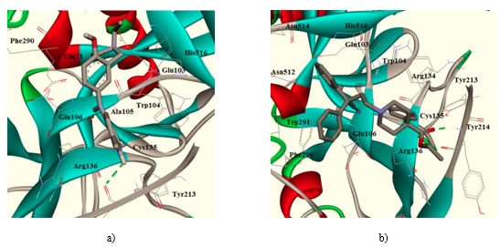
Figure 1. Hydrophobic and hydrogen bonding interactions of amodiaquine (a) and diphenoxylate (b) with amino acids in the GP receptor binding site.
The molecular docking analysis of ketotifen revealed that its primary interactions are hydrophobic, with only one hydrogen bond. This places ketotifen second in binding energy, behind diphenoxylate. Additionally, ketotifen has a rigid, inflexible structure due to its lack of rotatable bonds and its cyclic tertiary amine, which likely contributes to its reduced anti-Ebola activity compared to amodiaquine.
Among the drugs tested, only chloroquine, diphenoxylate, diphenylpyraline, and ketotifen demonstrated inhibitory activity against Ebola virus replication. Other compounds were either ineffective or untested. As VP40 is crucial for virus replication, its interaction with these drugs was explored. The docking simulations indicated that all four drugs formed hydrophobic interactions with the active-site hydrophobic pocket. Among these, chloroquine and diphenoxylate also formed hydrogen bonds with Lys212 and Lys127, respectively, whereas diphenylpyraline only formed hydrophobic interactions without hydrogen bonds.
Ketotifen showed the least favorable binding energy with the VP40 receptor. These findings underscore the importance of modifying certain parts of the drug to improve its binding properties by strengthening hydrophobic interactions or hydrogen bonding, thereby enhancing its potency. Structure-activity relationship (SAR) analysis showed that compounds with aromatic rings, hydrophobic groups, tertiary amines, rotatable bonds, and optimal hydrogen-bonding properties are more likely to exhibit stronger anti-Ebola activity. The key to increasing their anti-Ebola power lies in the right balance of hydrogen bonds and high lipophilicity, which boosts hydrophobic interactions. These compounds, therefore, represent potential candidates for the development of new Ebola treatments.
This study explored the potential of licensed non-viral drugs to inhibit Ebola virus entry and proliferation using bioinformatics. The analysis identified several key interactions between drugs and their respective receptors, including hydrophobic, π-π, hydrogen, and cation-π bonds. Among the drugs studied, chloroquine, diphenoxylate, and amodiaquine showed the most promising docking results, exhibiting the strongest binding affinities for the GP and VP40 proteins. In contrast, erythromycin and dirithromycin demonstrated weaker binding due to their high hydrophilicity. The results suggest that drugs with hydrophobic components, optimal hydrogen-bonding interactions, and tertiary amines have greater potential to inhibit Ebola virus activity. The findings propose these compounds as viable candidates for further development in the treatment of Ebola virus infections.
None
None
None
None
Open Access The author(s) retain copyright. This article is licensed under the Creative Commons Attribution-NonCommercial-ShareAlike 4.0 International License. It may be shared and adapted for non-commercial purposes with appropriate attribution, an indication of changes, and distribution of adaptations under the same license. Third-party material may be subject to separate terms identified in its credit line. View the license at https://creativecommons.org/licenses/by-nc-sa/4.0/.
 Publication System