The application of various agricultural pesticides can lead to adverse effects on bodily tissues. This study evaluated the impact of dermal exposure to malathion on sex hormone levels and its potential carcinogenicity in mice. 30 adult male mice were randomly assigned to control and treatment groups. The treatment group received a topical application of malathion at 1 ppm (1 ml) for 12 weeks. After the exposure period, serum levels of testosterone, luteinizing hormone (LH), and follicle-stimulating hormone (FSH) were analyzed, and tissue samples were collected post-necropsy. Cytotoxicity was assessed using the micronucleus assay and MTT method in vitro, and the IC50 value was calculated. Results showed no significant difference in LH levels between groups; however, both testosterone and FSH levels declined significantly in the treated mice. Histological evaluation revealed mild liver toxicity in the treatment group. Additionally, the frequency of micronuclei at various Malathion concentrations was significantly elevated compared to controls (P < 0.05). Overall, these findings suggest that dermal exposure to malathion can reduce reproductive hormone levels and induce cytotoxicity in mice.
Exposure to pesticides is a significant health concern. Organophosphates, a class of pesticides, include several compounds, with malathion among the most commonly used. These pesticides affect cholinesterase enzymes, reduce insulin secretion, disrupt the metabolism of proteins, carbohydrates, and fats, and cause cytotoxicity. They also impair mitochondrial function, leading to cellular oxidative stress and affecting the nervous and endocrine systems. Organophosphates are absorbed through the skin, respiratory tract, and digestive system and are rapidly metabolized into active forms [1-3].
Humans can come into contact with these toxic substances through various sources, including occupational exposure in agriculture, contaminated food, and environmental factors. Those living in agricultural areas are often exposed to higher levels of organophosphates, endotoxins, and allergens than the general population. As a result, a combination of these substances is linked to various health issues, including cancer and reproductive disorders. One key impact of organophosphates is their effect on sex hormones, as they can reduce hormone secretion by disrupting the endocrine system [4-7].
The effects of these toxins are dose-dependent and vary with exposure duration. Continuous exposure, especially in agricultural settings, may pose a risk to the reproductive health of both humans and animals. Despite numerous studies exploring the potential connection between organophosphates and increased risks of carcinogenesis and reproductive issues, the results remain inconclusive. While some research has examined the carcinogenic potential of malathion in humans, there is limited, conflicting evidence on this matter [8, 9], with no definitive or comprehensive studies conducted to date. This study aims to explore the toxic effects of malathion on fibroblast cells and liver tissues, as well as on the levels of sex hormones, including testosterone, in mice.
This study involved 30 adult male white mice, each weighing 20-30 grams and aged 10-12 weeks. The mice were randomly divided into two groups, each consisting of 15 animals: a control group and a treatment group. In the in vivo component of the experiment, the control group received only physiological saline. In contrast, the treatment group received 1 mL of a 1 ppm Malathion solution applied topically to the dorsal skin once daily for 12 weeks. The mice were housed in standard cages under controlled conditions at 25 °C with a 12-hour light/dark cycle, and all procedures were conducted in accordance with established ethical guidelines for animal research. Following the final administration, samples were collected from both groups.
Blood samples were drawn by general anesthesia, with approximately 3-4 ml collected from the axillary region into test tubes without anticoagulants. The mice were subsequently euthanized, and their tissues were harvested. The blood was centrifuged at 2000 rpm for 15 minutes, and the resulting serum was separated and stored at -70 °C in microtubes for subsequent analysis. Hormonal levels of testosterone, follicle-stimulating hormone (FSH), and luteinizing hormone (LH) were assessed by ELISA, in which serum samples and standards were applied to a 96-well plate. After the ELISA protocol, readings were taken at 450 nm using a spectrophotometer. Tissue samples were taken from various organs, fixed in 10% buffered formalin, and then prepared into histological sections for examination.
In vitro cytotoxicity of malathion was evaluated on L929 mouse fibroblast cells using both the MTT assay and the micronucleus test. Additionally, the morphological alterations in the cells due to Malathion exposure were observed and documented.
L929 fibroblast cells were cultured in 25 cc flasks using PRMI-1640 medium supplemented with 10% fetal calf serum (FCS), 100 units/ml penicillin, 100 μg/ml streptomycin, 2 mM L-glutamine, and 5.12 μM HEPES. Cells were cultured at 37 °C and 5% CO2, passaged until reaching the optimal growth stage (typically after 3-4 passages). The cells were detached from the flasks using EDTA and assessed for viability through the Trypan blue exclusion method. The cells were seeded in six-well plates at 5 × 10^4 cells/ml and exposed to malathion at concentrations of 10, 25, 50, and 100 µg/ml. After 48 hours, cell morphology was examined for attachment, granularity, and other changes using an inverted light microscope and digital imaging.
The cytotoxicity of malathion was assessed using the MTT assay to evaluate cell proliferation. The growth inhibition percentage was calculated from optical absorbance readings obtained with the ELISA reader.
The IC50 value was determined by constructing a dose-response curve using various concentrations of the toxin and the corresponding percentage of viable cells.
This test used the binucleate cell method during cytokinesis, as described by Fenech. After incubating the cells with malathion at varying concentrations for 24 hours, the cells were collected. Cells were separated using 25% trypsin and then centrifuged at 1000 rpm for 10 minutes. The cell pellet was treated with a fixative solution (methanol:acetic acid, 8:1). The resulting suspension was spread onto glass slides and allowed to air-dry. The slides were stained with 10% Giemsa solution for 7 minutes. Cell counting was carried out using an Olympus BH2 microscope at 1000x magnification. Between 300 and 700 binucleate cells were counted per slide. The percentage of binucleate cells containing micronuclei was calculated using a specific formula, and the total number of binucleate cells was also recorded.
The statistical analysis was conducted using a one-way analysis of variance (ANOVA). Since the Kolmogorov-Smirnov test indicated a normal distribution, parametric tests were employed. Duncan’s post-hoc test was applied to identify which groups differed significantly when the ANOVA showed a significant result. All statistical computations were carried out using SPSS version 23, with a significance threshold of 0.05. Data are presented as Mean ± Standard Deviation. Graphs were created with Excel, and IC50 values were determined using Prism software.
Histopathological analysis was performed using hematoxylin-eosin (H & E) staining to assess liver tissue changes relative to normal tissue (Figure 1). The parameters examined included tumor formation, cytotoxicity, and necrosis, as well as conditions such as Hepatitis, Cholestasis, and Steatosis. The treated group exhibited mild hepatotoxic effects.
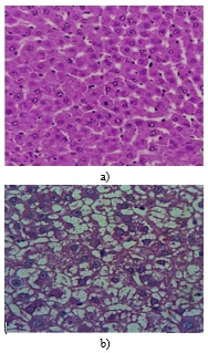
Figure 1. The impact of malathion on liver tissue observed under 4x magnification, stained with hematoxylin-eosin: (a) Liver tissue from the control group showing normal structure; (b) Presence of fat accumulation in liver cell cytoplasm and nuclear pyknosis in liver cells due to malathion exposure.
When different concentrations of malathion (10, 25, 50, and 100 μg/mL) were tested on L929 mouse fibroblast cells, a noticeable inhibition of cell growth was observed. The highest inhibition rates were observed at 50 μg/ml (27.19%) and 100 μg/ml (30.24%) (Table 1).
Table 1. Effect of malathion concentrations on inhibition of L929 mouse fibroblast cell growth after 24 hours.
Concentration (μg/ml) | Average Optical Absorbance ± Standard Deviation | Average Inhibition Percentage |
10 | 0.09 ± 0.01 | 5.5% |
25* | 0.04 ± 0.07 | 20.37% |
50* | 0.03 ± 0.002 | 27.19% |
100* | 0.032 ± 0.021 | 30.24% |
Control | 0.1 ± 0.01 | - |
Significant difference compared to the control group (P < 0.05).
The optical absorbance at concentrations of 25, 50, and 100 μg/ml was significantly different from the control group, indicating a notable inhibition of cell growth (P < 0.05). The IC50 for Malathion in this cell line was 22.90 μg/ml after 24 hours of treatment.
The frequency of micronuclei in cells treated with malathion at various concentrations was measured and showed no significant differences compared to the control group (Table 2).
Table 2. Frequency of micronuclei in cells exposed to malathion.
Concentration (μg/ml) | Frequency of micronucleus (%) |
Control | 3.04% |
10 | 3.62% |
25 | 3.85% |
50 | 3.65% |
100 | 4.04% |
The study found a significant reduction in the levels of testosterone and FSH hormones in the treatment group compared to the control group, suggesting that malathion disrupted the hormonal balance in the body. Although a similar decrease in LH levels was observed, it was not statistically significant (Table 3).
Table 3. Effect of malathion on LH, FSH, and testosterone concentrations in mice.
Hormone (concentration) | Control group | Treatment group |
FSH (IU/L)* | 280% | 103% |
LH (IU/L) | 320% | 100% |
Testosterone (ng/dL)* | 30.25 | 5.50 |
Significant difference compared to the control group (P < 0.05).
The results demonstrate that different Malathion concentrations significantly inhibit fibroblast growth in vitro. Skin contact with malathion reduced blood levels of FSH and testosterone in mice. Histological examination confirmed mild hepatotoxicity in the treated group. Previous studies have shown that malathion exposure increases breast cancer incidence in rats and is associated with liver, nasal, and oral tumors following long-term exposure. Furthermore, malathion has been linked to kidney irritation in laboratory animals. Despite limited research on malathion’s liver effects in agricultural workers, existing evidence suggests that malathion exposure can cause genetic damage and has been associated with various cancers, including breast cancer and non-Hodgkin lymphoma in farmers [10-20].
Malathion is quickly absorbed through the skin, with the extent of absorption depending on both the dose and the area exposed [16]. In one toxicokinetic study, rats that received an oral dose of 28 mg/kg and a dermal dose of 41 mg/kg of malathion absorbed over 90% of the chemical, which was then excreted in their urine within 24 hours. Traces of Malathion were also found in their feces, kidneys, blood, liver, and intestines. Another study showed that inhalation exposure to malathion for two weeks caused its accumulation in the liver and kidneys, resulting in toxicity in these organs [17].
Research has demonstrated that malathion is mutagenic, with studies showing genetic damage in various species, including bacteria, fruit flies, hamsters, mice, and fish. The National Institute for Occupational Safety and Health (NIOSH) recognizes malathion as a mutagen. In vitro studies have confirmed that malathion induces genetic damage when ingested by mice [18].
Long-term oral exposure to malathion in rats has been linked to an increase in the incidence of tumors, including those in the oral cavity, nasal passages, and liver. However, in an 80-week study in which rats were given daily doses of 0, 359, or 622 mg/kg of malathion, no significant carcinogenic effects were observed [19]. Similarly, a study that examined dietary exposure to 0, 166, and 332 mg/kg/day of malathion for 103 weeks in rats also did not show any carcinogenic effects [21]. However, a two-year study with female mice, who were given doses ranging from 2 to 868 mg/kg per day, found a significant increase in liver tumors, including adenomas and carcinomas, at the highest doses tested [22].
In a rat bioassay, when malathion was administered at doses of 1476 and 2978 mg/kg/day in males and 1707 and 3448 mg/kg/day in females, it induced liver adenomas and hepatocellular carcinoma, supporting its carcinogenicity [23]. The Environmental Protection Agency (EPA) classified malathion as a possible carcinogen in 1999, though a detailed dose-response evaluation has yet to be performed. The Federal Insecticide, Fungicide, and Rodenticide Act (FIFRA) also lists malathion as a potential carcinogen.
In a 2003 study, rats were exposed to malathion for 4 hours daily over 28 days. The results showed that doses of 8 and 16 mg did not affect the rats’ internal organs [24]. Another investigation looked at the non-cholinergic effects of malathion on apoptosis in L929 mouse fibroblasts. The study demonstrated that malathion promotes apoptosis in a dose- and time-dependent manner, as assessed by flow cytometry and caspase activation [25].
A 2012 study on the cytogenetic toxicity of malathion in rats found that exposure to the chemical resulted in a significant increase in chromosomal abnormalities and DNA damage, suggesting that malathion is both genotoxic and clastogenic [26]. Studies of workers exposed to malathion also showed an increase in chromosomal abnormalities, with a dose-dependent effect observed [27].
A risk assessment of workers exposed to organophosphorus pesticides, including malathion, revealed an increase in chromosomal abnormalities such as acentric fragments and micronuclei [28].
In our study, while a dose-dependent increase in micronuclei was observed following Malathion exposure, it was not statistically significant. This result is consistent with another study examining the genotoxic effects of Malathion on HTC rat cells, which reported no significant increase in micronuclei at concentrations of 9, 0.009, and 0.0009 mg [29].
Our findings also showed a significant decrease in testosterone and FSH hormone levels in the treatment group compared to the control group, suggesting that malathion disrupts the hormonal system. Although a similar reduction in LH levels was noted, it was not statistically significant. A separate study investigating the protective effects of green tea extract on ovarian tissue function in Malathion-treated rats found that malathion negatively affected sex hormone secretion and oogenesis in female rats. However, the green tea extract helped mitigate these adverse effects [30].
The impact of malathion on sex hormones and potential skin carcinogenesis was assessed in mice. The findings revealed that while luteinizing hormone levels were unaffected, significant reductions in follicle-stimulating hormone and testosterone were observed. Additionally, histological analysis showed mild liver damage in the treatment groups. Malathion exposure also caused a notable increase in micronuclei frequency compared to controls. These results suggest that skin exposure to malathion may disrupt hormonal balance and cause potential harm to body tissues.
None
None
None
None
Open Access The author(s) retain copyright. This article is licensed under the Creative Commons Attribution-NonCommercial-ShareAlike 4.0 International License. It may be shared and adapted for non-commercial purposes with appropriate attribution, an indication of changes, and distribution of adaptations under the same license. Third-party material may be subject to separate terms identified in its credit line. View the license at https://creativecommons.org/licenses/by-nc-sa/4.0/.
 Publication System