Gout disease is recognized as one of the most prevalent forms of arthritis worldwide, with an estimated prevalence of around 2.5%. Its pathophysiology is primarily linked to elevated levels of uric acid in the bloodstream, which may result from either increased production or reduced renal excretion. Clinically, it often presents as a swollen, erythematous, and painful joint. The definitive method for diagnosis remains synovial aspiration from the affected joint. While acute attacks typically require management in the emergency department, long-term care is generally provided in primary care settings.
This literature review aims to explore gout disease in terms of its clinical features, diagnostic approaches, and treatment strategies, particularly focusing on the role of primary care. A comprehensive search was conducted on the PubMed database using the MeSH term “gout disease” to identify pertinent studies. Gout disease continues to be among the most widespread rheumatologic conditions globally, and unhealthy lifestyle choices, including alcohol intake and high consumption of red and white meats, may exacerbate its incidence. Diagnosis may involve blood tests, radiographic imaging, and joint aspiration. Maintenance therapy, often administered in primary care, includes the use of Allopurinol or other urate-lowering agents.
Gout is recognized as the most common and widespread form of arthritis (joint inflammation), and among all arthritis-related conditions, it holds the most significant potential for effective and safe treatment [1–3]. The primary cause of this disease is elevated uric acid levels in the body, which can result from either increased synthesis or reduced renal clearance. Management strategies target these mechanisms by using xanthine oxidase inhibitors to lower production or by enhancing renal excretion of uric acid [4–6]. This review provides a brief overview of the disease’s epidemiology, pathophysiology, and therapeutic options, with a particular emphasis on gout management within primary care settings.
This literature review aims to explore gout disease in terms of its clinical features, diagnostic approaches, and treatment strategies, particularly focusing on the role of primary care. A comprehensive search was conducted on the PubMed database using the MeSH term “gout disease” to identify pertinent studies.
As highlighted earlier, gout disease is among the most frequently encountered conditions in clinical practice, affecting roughly 2.5% of the general population. The condition is notably more common in men than in women, with estimates suggesting a prevalence of 3%–6% in males and 1%–2% in females. In recent years, the number of gout cases has increased significantly, reaching prevalence rates of up to 10% in some countries. This upward trend is attributed mainly to unhealthy lifestyle choices, such as increased consumption of junk food and decreased physical activity, both of which play a major role in the development of the disease [7].
Urate, the ionized form of uric acid, begins to crystallize and deposit in tissues when serum uric acid levels exceed the critical threshold of 6.8 mg/dL [8]. These urate deposits may arise from either overproduction of uric acid—accounting for roughly 10% of cases—or from impaired renal excretion, which is the predominant cause, responsible for approximately 90% of gout disease [9]. Several factors contribute to elevated uric acid production, including:
· Enzymatic disorders such as Lesch-Nyhan syndrome are X-linked recessive conditions. In this syndrome, a deficiency in a purine metabolism enzyme leads to excessive uric acid accumulation [10].
· Dietary habits play a significant role in raising uric acid levels. Uric acid is commonly elevated by diets high in animal proteins—particularly red and white meat. Therefore, individuals with gout are advised to limit meat intake, increase their consumption of vegetables, and avoid alcohol, as it is a well-known contributor to elevated uric acid levels [11].
Elevated uric acid levels can arise from internal (endogenous) sources such as malignancies or chemotherapy, both of which increase cellular turnover and elevate purine concentrations in the body [12]. However, the predominant cause—accounting for about 90% of gout disease—is decreased renal excretion of uric acid [8]. Because uric acid is poorly soluble, its elimination requires specific transporters, such as URAT1 (uric acid transporter 1) and OAT (organic anion transporter). Usually, around 10% of uric acid is excreted through the urine. In cases involving uromodulin gene mutations, this excretion rate drops significantly, leading to elevated serum uric acid levels due to impaired uric acid elimination [13].
Gout presents in both acute and chronic forms, each requiring different management approaches. In acute gout, uric acid crystals activate synovial macrophages, which attempt to engulf these crystals. While mature macrophages can phagocytose urate crystals without eliciting inflammation, immature macrophages may initiate an immune response by releasing inflammatory cytokines and activating mast cells and neutrophils. Treatment during acute flare-ups typically includes NSAIDs (non-steroidal anti-inflammatory drugs) or colchicine, both of which help suppress the inflammatory process [14].
Chronic gout results from repeated acute episodes, and persistent recurrence is a hallmark of the condition [15]. Research indicates that chronic gout is associated with chondrocyte activation, leading to increased production of inflammatory mediators and elevated matrix metalloproteinase (MMP) levels, which contribute to cartilage degradation [16]. Even when acute gout is well-managed, chronic gout may still develop [8]. However, due to advancements in healthcare, the incidence of chronic gout has declined. Long-term treatment generally involves Allopurinol, a xanthine oxidase inhibitor, while NSAIDs and Colchicine are usually discontinued once inflammation is controlled [17].
In acute gout, the clinical signs often include joint pain and swelling, particularly affecting areas such as the metatarsophalangeal joint, knee, foot, olecranon, and pre-patellar bursa. Symptoms include erythema, joint swelling, stiffness, and pain, often triggered by infection, trauma, or dehydration (Figure 1) [18]. Chronic gout is driven by repeated acute episodes, resulting in the accumulation of monosodium urate (MSU) crystals as firm, white-to-yellow subcutaneous deposits with a heterogeneous texture. These are typically accompanied by chronic joint stiffness and persistent pain.
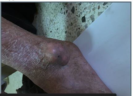
Figure 1. Gouty arthritis.
The definitive diagnosis of gout relies on joint aspiration followed by examination of the synovial fluid using polarized light microscopy, although this procedure is invasive [19]. Key conditions that need to be differentiated from gout include septic arthritis, pseudo-gout, and reactive arthritis, which must be excluded to establish an accurate diagnosis [4]. Blood tests often reveal elevated neutrophil counts, increased inflammatory markers, and hyperuricemia; however, these findings alone are insufficient to confirm gout [20].
Aspiration of synovial fluid is a reliable method to exclude septic arthritis by culturing the fluid, and the presence of monosodium urate (MSU) crystals can be confirmed through polarized light microscopy (Figure 2) [21]. Measuring urinary uric acid excretion is generally not useful beyond the early stages of the disease; however, a 24-hour collection to assess the uric acid-to-creatinine ratio may be performed [19]. Among imaging modalities, ultrasonography is highly sensitive for detecting MSU crystal deposits. It demonstrates the characteristic “double contour sign,” which indicates urate crystal deposition on the surface of hyaline cartilage; however, this imaging technique is not entirely specific (Figure 3) [22].
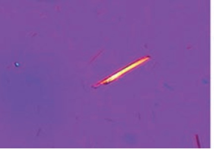
Figure 2. Monosodium urate crystal under polarised light microscopy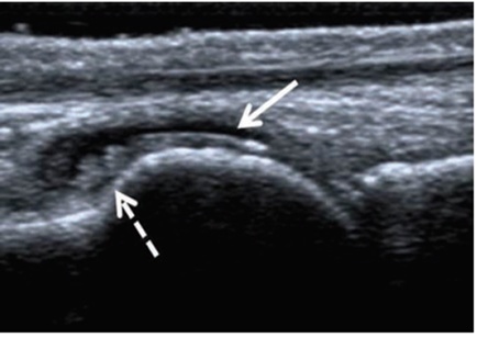
Figure 3. Radiographic appearance of gouty arthritis.
The 2017 guidelines from the British Society of Rheumatology and the British Health Professionals in Rheumatology recommend NSAIDs or low-dose colchicine (500 mcg, taken 2 to 4 times daily) as the first-line treatment for acute gout attacks, tailored to the patient’s individual profile and preferences. For patients who cannot use NSAIDs or colchicine due to contraindications, a low dose of prednisolone (35 mg daily for five days) is advised. Strict adherence to short-term treatment is essential to reduce the risk of recurrence and prevent progression to chronic gout. Lifestyle changes are also critical for gout patients; they should avoid alcohol consumption and limit their intake of foods high in purines, such as red and white meat [23].
Patients with gout should engage in more physical activity due to their elevated risk for cardiovascular disease and diabetes mellitus. They must also be mindful of medications that can increase uric acid levels, such as thiazide and loop diuretics, which are best avoided in gout cases; alternative antihypertensive therapies should be considered instead. In terms of urate-lowering therapy (ULT), Allopurinol—a xanthine oxidase inhibitor—is the preferred and most effective option. Current guidelines recommend starting ULT for all patients proactively, without waiting for complications or frequent gout flare-ups. Furthermore, patients must receive clear instructions on medication usage, with the treatment regimen simplified and the therapy’s goals thoroughly explained [23].
Moreover, neglecting the treatment regimen can lead to complications, highlighting the importance of strict adherence. Guidelines recommend starting Allopurinol at a dose of 50-100 mg per day, with gradual increments of 50-100 mg every four weeks until the serum uric acid level reaches the target. According to British guidelines, this optimal serum uric acid concentration is 300 micromol/L. In contrast, European and American guidelines set the target at 360 micromol/L. The maximum daily dose of Allopurinol can be up to 900 mg, divided into multiple doses. Recent studies suggest that excessively low serum uric acid levels may increase the risk of dementia and Parkinsonism, so extremely low levels should be avoided. Additionally, guidelines advise using 500 mcg of colchicine as prophylaxis for six months to reduce the chance of gout recurrence. For those who cannot tolerate Colchicine, NSAIDs combined with gastroprotective agents are recommended to prevent ulcers during prophylactic treatment [23].
Gout affects approximately 2.5% of the population, with prevalence rates reaching up to 10% in certain countries due to the rise in unhealthy habits such as consuming fast food and a lack of physical activity. The primary cause of gout is the elevation of uric acid or urate crystals, which results from an imbalance between uric acid production and its elimination. Therefore, the primary focus of gout treatment involves lowering uric acid levels using Allopurinol, while managing acute flare-ups with NSAIDs and Colchicine, with the latter also playing an essential role in prophylactic therapy to prevent recurrent attacks.
None
None
None
None
Open Access The author(s) retain copyright. This article is licensed under the Creative Commons Attribution-NonCommercial-ShareAlike 4.0 International License. It may be shared and adapted for non-commercial purposes with appropriate attribution, an indication of changes, and distribution of adaptations under the same license. Third-party material may be subject to separate terms identified in its credit line. View the license at https://creativecommons.org/licenses/by-nc-sa/4.0/.
 Publication System