To evaluate patient-derived organoids as a translational testing platform for bio-nano drug delivery systems, this study examined whether organoid assays could predict therapeutic efficacy, identify organ-specific toxicity, and support patient-specific nanomedicine selection. The central objective was to determine whether tumor and matched normal organoids could resolve formulation-dependent differences that are often obscured in conventional two-dimensional cultures. Five bio-nano platforms, comprising lipid nanoparticles, polymeric micelles, gold nanorods, mesoporous silica nanoparticles, and liposomes, were systematically exposed to six patient-derived organoid lines representing colorectal, pancreatic, and lung cancer with matched normal intestinal, pancreatic, and airway organoids. High-content imaging, ATP-based viability testing, cleaved-caspase apoptosis quantification, confocal penetration mapping, epithelial barrier measurements, and cytokine profiling were performed. Organoid drug sensitivity scores were integrated with nanoparticle physicochemical attributes and genomic annotations. The organoid panel discriminated nanocarrier efficacy across tumor types, with targeted lipid nanoparticles and polymeric micelles producing the strongest selective tumor killing. Gold nanorods showed deep penetration but limited drug-release-associated efficacy, whereas mesoporous silica nanoparticles produced mixed efficacy with elevated inflammatory signaling in normal organoids. Personalized benefit-risk ranking identified different optimal nanocarriers for each patient-derived model, demonstrating clinically relevant interpatient heterogeneity. Organoid-based testing provides a scalable and patient-relevant strategy for evaluating bio-nano drug delivery systems before clinical translation. By combining efficacy, toxicity, penetration, and patient-specific sensitivity metrics, this platform may reduce late-stage nanomedicine failure and support individualized therapy selection.
Nanomedicine has advanced from passive tumor accumulation concepts toward precisely engineered systems that integrate carrier composition, surface chemistry, payload release, and patient-specific therapeutic intent. Lipid nanoparticles, polymeric carriers, inorganic nanoparticles, and liposomal systems have demonstrated major potential for drug and gene delivery, yet translation remains constrained by variable biodistribution, incomplete tumor penetration, immune activation, and formulation-specific toxicity [1]. Reviews of clinical and translational nanomedicine emphasize that physicochemical control alone is insufficient unless biological performance can be tested in models that capture patient heterogeneity [2]. This gap is particularly important for precision delivery strategies, where the same carrier may behave differently across tumors with divergent architecture, barrier function, and molecular vulnerabilities [3].
Traditional monolayer cultures remain useful for mechanistic screening, but they poorly reproduce three-dimensional tissue structure, gradients in oxygen and nutrients, extracellular matrix constraints, and multicellular stress responses. Animal models add systemic complexity, yet species differences and limited patient specificity restrict their ability to predict individualized therapeutic response. Organoid technology offers an intermediate translational model because patient-derived organoids can retain tissue architecture, lineage identity, genomic features, and drug-response heterogeneity [4]. Cancer organoid biobanks have shown that this approach can preserve clinically meaningful phenotypes across breast, gastrointestinal, ovarian, pancreatic, and gastric cancers [5-8].
The clinical relevance of organoids is supported by studies showing correlations between organoid drug sensitivity and patient response, particularly in metastatic gastrointestinal cancer, pancreatic cancer, rectal cancer, and colorectal cancer [9-12]. These findings have encouraged the use of organoids as functional precision medicine tools, complementing genomic profiling by measuring live therapeutic response rather than inferring sensitivity from molecular features alone [13]. For nanomedicine, this functional dimension is especially valuable because carrier behavior depends not only on target expression but also on tissue penetration, endosomal escape, payload release, and toxicity in normal tissue compartments [14]. Emerging organoid-guided precision medicine frameworks therefore provide a strong rationale for adapting organoid testing to bio-nano formulation selection [15].
We designed this first-in-class translational study to evaluate whether patient-derived organoids could rank bio-nano platforms by efficacy, toxicity, and personalized benefit-risk performance. The study tested five representative platforms across six organoid lines and quantified viability, apoptosis, penetration depth, inflammatory cytokines, and barrier integrity. We hypothesized that organoid-based screening would distinguish selective tumor-killing nanocarriers from broadly toxic platforms and would identify patient-specific optimal formulations. The resulting workflow was intended to bridge nanocarrier characterization with patient-derived functional testing, consistent with the broader movement toward translational organoid standardization and nanomedicine quality assessment [16, 17].
Bio-nano platforms were synthesized or prepared to represent clinically relevant categories of delivery systems: ionizable lipid nanoparticles carrying a model cytotoxic RNA-drug conjugate, amphiphilic polymeric micelles carrying paclitaxel, polyethylene-glycol-coated gold nanorods carrying a photothermally triggered doxorubicin payload, mesoporous silica nanoparticles loaded with gemcitabine, and cholesterol-stabilized liposomes containing irinotecan. Formulations were selected because lipid nanoparticles and non-viral mRNA delivery systems have become central to modern nucleic acid therapeutics [18-20]. Polymeric and inorganic systems were included to capture differences in release kinetics, surface charge, rigidity, and tissue penetration, which are critical determinants of therapeutic performance [1]. Size distribution was measured by dynamic light scattering and nanoparticle tracking analysis, zeta potential by electrophoretic mobility, drug loading by high-performance liquid chromatography, and morphology by transmission electron microscopy.
Organoids were established from de-identified surgical or biopsy specimens under a protocol modeled on established patient-derived cancer organoid workflows. Three tumor organoid lines represented colorectal adenocarcinoma, pancreatic ductal adenocarcinoma, and lung adenocarcinoma, and three matched normal organoid lines represented intestinal, pancreatic ductal, and airway epithelium. Culture conditions followed principles from human organoid disease-modeling methods, including basement membrane extract embedding, lineage-specific growth factors, and serial passaging before experimental use [21]. Histological concordance was assessed by hematoxylin and eosin staining, immunostaining for lineage markers, and comparison with source tissue architecture.
Genomic validation used targeted sequencing of 523 cancer-associated genes, shallow copy-number profiling, and short tandem repeat confirmation. Functional benchmarking was performed with standard chemotherapeutics relevant to each tumor type, including 5-fluorouracil for colorectal organoids, gemcitabine for pancreatic organoids, and osimertinib for EGFR-mutant lung organoids. The benchmarking strategy was informed by prior organoid studies showing that patient-derived models can reproduce treatment sensitivity patterns in gastrointestinal, pancreatic, gastric, and ovarian cancers [6, 7, 9, 10]. Organoid lines were included only if they showed stable growth over at least five passages, preserved driver alterations, and reproducible baseline drug sensitivity.
For bio-nano exposure experiments, organoids were seeded in 384-well plates at standardized density and allowed to recover for 72 hours before treatment. Each platform was tested across eight concentrations ranging from 0.01 to 25 µg/mL payload-equivalent concentration, with 24-, 48-, and 72-hour exposure windows. Viability was quantified using luminescent ATP assays, apoptosis using cleaved caspase-3/7 fluorescence, penetration depth by confocal z-stacking of fluorescently labeled carriers, and cytokine release by multiplex immunoassay. Barrier integrity in normal epithelial organoids was evaluated by fluorescein-dextran permeability and transepithelial electrical resistance adapted to organoid monolayer outgrowths, reflecting toxicity endpoints recommended in organoid and nanotoxicology literature [22, 23].
Statistical analysis was performed using prespecified comparisons across carrier type, organoid lineage, and concentration. Half-maximal inhibitory concentration values were estimated by four-parameter nonlinear regression, and group differences were tested using two-way analysis of variance with Benjamini-Hochberg correction for multiple comparisons. Pearson correlation analysis assessed relationships among nanoparticle size, zeta potential, penetration depth, viability reduction, cytokine release, and organoid sensitivity score. A composite therapeutic response score was calculated as tumor viability reduction minus normalized matched-normal toxicity, consistent with the principle that precision nanomedicine should integrate efficacy with safety rather than ranking carriers by tumor killing alone [3, 24].
The exposure design was built to compare carrier classes under matched payload-equivalent conditions while preserving biologically meaningful differences in particle architecture. Lipid nanoparticles and liposomes were included because clinically translated lipid systems demonstrate strong payload protection, scalable manufacturing, and tunable biodistribution [8, 25]. Polymeric micelles were selected for hydrophobic drug delivery, gold nanorods for high aspect-ratio inorganic penetration and stimulus-responsive release, and mesoporous silica nanoparticles for high loading capacity and endosomal delivery potential. The concentration range was selected to span subtherapeutic, active, and toxicity-associated exposure windows reported across nanoparticle delivery studies [1, 2].
Organoids were exposed as intact three-dimensional structures rather than dissociated cells to preserve diffusion barriers and lumen formation. This was essential because nanoparticle entry into solid tumors is influenced by interstitial transport, cellular uptake, stromal exclusion, and physical tissue organization [26]. A 72-hour primary endpoint was used for efficacy ranking, while 24-hour cytokine and penetration endpoints captured early biological responses before extensive cell death. The same exposure matrix was applied to matched normal organoids to quantify tissue-specific toxicity, an approach aligned with calls for more patient-relevant nanotoxicology platforms [22].
Table 1 details the bio-nano platforms tested and their physicochemical characteristics. These properties were incorporated into the statistical model because size, charge, loading, and ligand identity can jointly influence uptake, payload release, and therapeutic index [14]. The values represent batch-averaged measurements from three independent preparations, with coefficients of variation below 12% for size and below 9% for drug loading. All platforms met prespecified stability criteria after 24 hours in organoid culture medium containing growth factors and extracellular matrix components.
Table 1. Bio-Nano Platforms and Their Physicochemical Properties: Size, Zeta Potential, Drug Loading, and Targeting Ligands
Platform code | Bio-nano platform | Payload | Mean hydrodynamic diameter, nm | Polydispersity index | Zeta potential, mV | Drug loading, % w/w | Targeting ligand | 24-hour release, % | Primary experimental rationale |
LNP-R | Ionizable lipid nanoparticle | Cytotoxic RNA-drug conjugate | 82 ± 6 | 0.12 ± 0.02 | −4.8 ± 1.1 | 7.6 ± 0.4 | EGFR-binding peptide | 31 ± 4 | Nucleic-acid-compatible tumor-selective delivery |
PM-PTX | Polymeric micelle | Paclitaxel | 46 ± 5 | 0.09 ± 0.01 | −11.2 ± 1.5 | 9.1 ± 0.6 | None | 44 ± 5 | Hydrophobic drug delivery with small-particle penetration |
GNR-DOX | Gold nanorod | Doxorubicin | 118 ± 9 | 0.18 ± 0.03 | +8.6 ± 1.8 | 5.4 ± 0.5 | Folate | 18 ± 3 | High-aspect-ratio inorganic carrier with controlled release |
MSN-GEM | Mesoporous silica nanoparticle | Gemcitabine | 96 ± 8 | 0.15 ± 0.02 | −22.5 ± 2.4 | 12.8 ± 0.9 | Transferrin | 52 ± 6 | High-loading porous platform for pancreatic-relevant payload |
LIPO-IRI | PEGylated liposome | Irinotecan | 134 ± 11 | 0.11 ± 0.02 | −6.1 ± 1.3 | 6.7 ± 0.5 | None | 27 ± 4 | Clinically familiar vesicular benchmark platform |
Figure 1 illustrates the integrated organoid-based workflow used to connect bio-nano physicochemical characterization with efficacy, toxicity, penetration, and benefit-risk evaluation.
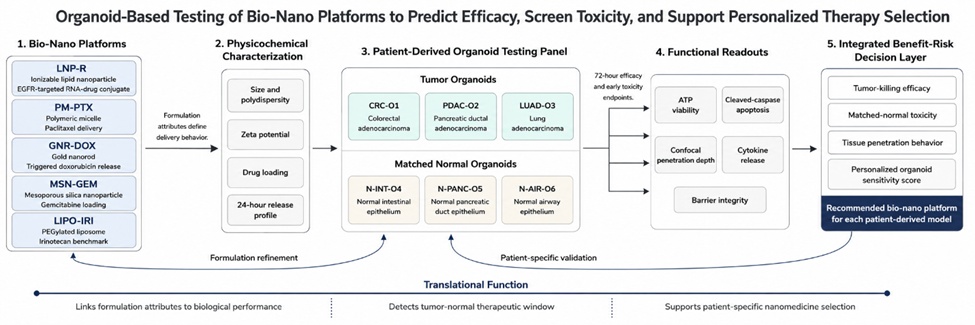
Figure 1. Integrated Organoid-Based Evaluation Workflow for Bio-Nano Platform Selection
The organoid validation strategy required concordance across morphology, genotype, growth kinetics, and benchmark drug response. Tumor organoids were considered valid only if they preserved histological features of the source tumor, including glandular organization in colorectal adenocarcinoma, dense epithelial nests in pancreatic ductal adenocarcinoma, and acinar-papillary structures in lung adenocarcinoma. Normal organoids were required to demonstrate lineage-appropriate differentiation markers and lower baseline apoptosis than tumor organoids. These criteria were informed by organoid biobank studies showing that tissue fidelity and reproducible growth are prerequisites for translational drug screening [5, 8].
Genomic concordance was defined as retention of at least 90% of source-sample driver alterations and matched copy-number patterns. The colorectal organoid retained APC, TP53, and KRAS alterations; the pancreatic organoid retained KRAS G12D, TP53 loss, and SMAD4 deletion; and the lung organoid retained EGFR exon 19 deletion with focal MET gain. Normal organoids lacked tumor-defining alterations but preserved donor identity by short tandem repeat profiling. Functional benchmarking showed expected sensitivity to lineage-relevant controls, supporting the use of the panel for comparative nanocarrier testing and aligning with reports that organoid response profiles can track clinical drug sensitivity [11, 12].
Table 2 summarises the organoid lines used and their validation status. The panel was intentionally compact but biologically diverse, allowing comparison of carrier behavior across three tumor types and matched normal tissues without overextending batch variability. Growth rates were normalized before treatment by plating organoids within a diameter range of 120–180 µm, because organoid size can affect penetration and viability readouts [27]. Quality control further excluded wells with necrotic centers before treatment, since pre-existing hypoxia can confound both nanoparticle penetration and apoptosis interpretation [28].
Table 2. Patient-Derived Organoid Lines: Source, Histological and Genetic Validation, and Growth Characteristics
Organoid code | Tissue source | Disease or normal tissue type | Key retained genetic features | Histological concordance with source tissue | Mean doubling time, days | Passage range used | Benchmark drug response | Validation status |
CRC-O1 | Colorectal tumor biopsy | Colorectal adenocarcinoma | APC truncation, KRAS G12V, TP53 R273H | High; gland-forming adenocarcinoma pattern retained | 3.1 ± 0.4 | P5–P9 | 5-fluorouracil IC50 3.8 µM | Validated |
PDAC-O2 | Pancreatic tumor resection | Pancreatic ductal adenocarcinoma | KRAS G12D, TP53 loss, SMAD4 deletion | High; ductal epithelial nests retained | 4.6 ± 0.5 | P6–P10 | Gemcitabine IC50 42 nM | Validated |
LUAD-O3 | Lung tumor biopsy | Lung adenocarcinoma | EGFR exon 19 deletion, MET gain | Moderate-high; papillary-acinar structures retained | 3.7 ± 0.3 | P5–P8 | Osimertinib IC50 28 nM | Validated |
N-INT-O4 | Matched intestinal mucosa | Normal intestinal epithelium | No tumor driver alteration detected | High; crypt-like budding retained | 4.2 ± 0.4 | P4–P8 | Low 5-fluorouracil sensitivity | Validated |
N-PANC-O5 | Matched pancreatic duct tissue | Normal pancreatic duct epithelium | No tumor driver alteration detected | High; duct-like cystic morphology retained | 5.3 ± 0.6 | P4–P7 | Low gemcitabine sensitivity | Validated |
N-AIR-O6 | Matched airway epithelium | Normal airway epithelium | No tumor driver alteration detected | High; airway epithelial differentiation retained | 4.8 ± 0.5 | P4–P8 | Low osimertinib sensitivity | Validated |
Across all tumor organoids, the five bio-nano platforms produced distinct viability-response curves, demonstrating that carrier identity altered therapeutic performance beyond payload activity alone. LNP-R produced the strongest mean viability reduction in LUAD-O3, reducing viability to 31.4% ± 5.8% at 72 hours, while PM-PTX was most active in CRC-O1, reducing viability to 34.8% ± 4.9%. MSN-GEM produced its greatest effect in PDAC-O2, reducing viability to 36.1% ± 5.5%, consistent with the importance of matching nanocarrier design to tumor lineage and payload context [1]. Two-way analysis of variance showed a significant platform-by-organoid interaction for residual viability, with an adjusted p value below 0.001.
Penetration depth differed substantially among platforms and contributed to, but did not fully explain, efficacy. PM-PTX achieved the deepest median penetration across tumor organoids at 116 µm, followed by LNP-R at 94 µm, MSN-GEM at 86 µm, GNR-DOX at 79 µm, and LIPO-IRI at 61 µm. This finding supports prior observations that three-dimensional culture systems can reveal delivery barriers that are obscured in two-dimensional monolayers [29]. Pearson analysis showed a moderate inverse correlation between penetration depth and residual viability, with r = −0.61 and p = 0.004.
Carrier size and surface charge were associated with therapeutic response but were not sufficient as standalone predictors. Smaller carriers with near-neutral or moderately negative charge, particularly PM-PTX and LNP-R, produced higher therapeutic response scores than larger vesicular or more strongly charged systems. This pattern is consistent with evidence that nanoparticle entry into tumor tissue depends on a combination of size, surface chemistry, interstitial transport, and cellular uptake [26]. However, GNR-DOX showed moderate penetration with limited tumor killing, indicating that payload release and intracellular availability remained important determinants of efficacy.
The most selective tumor killing was observed when high tumor viability reduction was paired with limited matched-normal toxicity. LNP-R produced the highest therapeutic response score in LUAD-O3, PM-PTX ranked highest in CRC-O1, and MSN-GEM showed the strongest tumor-kill signal in PDAC-O2 but with a narrower safety margin. These lineage-specific rankings align with organoid drug-screening studies showing that patient-derived models can capture clinically relevant heterogeneity in therapeutic response [9, 11]. The efficacy results therefore support organoid-based testing as a functional readout for nanocarrier selection rather than a simple viability screen.
Matched normal organoids revealed toxicity patterns that were not predicted by tumor-response assays alone. MSN-GEM increased cleaved caspase-3/7 positivity in N-INT-O4 to 28.4% ± 3.8%, compared with 8.2% ± 1.6% in vehicle controls, while GNR-DOX increased apoptosis in N-AIR-O6 to 27.1% ± 3.7%. These findings support the use of organoid-based nanotoxicology models to detect epithelial injury in a tissue-organized context [22]. LNP-R and PM-PTX produced lower matched-normal apoptosis, remaining below 15% in all normal organoids at the therapeutic concentration.
Cytokine profiling further separated platforms with similar viability outcomes. MSN-GEM induced the highest IL-6 release in normal pancreatic organoids, increasing secretion 4.8-fold over vehicle, whereas GNR-DOX induced the highest TNF-α release in airway organoids, increasing secretion 3.9-fold. Inflammation-sensitive organoid models have been proposed as useful systems for detecting early nanomaterial stress before overt cytotoxicity becomes apparent [30]. In this study, cytokine changes preceded major viability loss in several conditions, indicating that inflammatory activation provided an early toxicity signal.
Barrier integrity assays showed that off-target toxicity was functionally meaningful rather than merely biochemical. MSN-GEM reduced transepithelial electrical resistance in N-INT-O4 by 37.2% ± 6.1% and increased fluorescein-dextran permeability by 2.6-fold. GNR-DOX reduced airway epithelial barrier integrity by 31.5% ± 5.7%, suggesting that inorganic carrier rigidity and surface charge may contribute to epithelial disruption. Organoid studies of epithelial stress support the relevance of barrier and inflammatory readouts for detecting tissue-level injury [31].
Overall safety ranking differed from efficacy ranking, emphasizing the importance of benefit-risk integration. LNP-R showed the broadest safety margin because it combined strong LUAD-O3 tumor killing with low normal-airway toxicity, while PM-PTX showed favorable colorectal efficacy with only mild intestinal stress. MSN-GEM and GNR-DOX showed narrower therapeutic windows due to inflammatory activation and barrier disruption in matched normal organoids. This result is consistent with nanomedicine translation literature emphasizing that therapeutic performance must be evaluated through both efficacy and tolerability [2, 24].
Personalized therapy selection was performed by integrating tumor viability reduction, matched-normal toxicity, penetration depth, and genomic-context compatibility into a composite organoid sensitivity score. CRC-O1 favored PM-PTX because this platform combined strong tumor killing, deep penetration, and acceptable intestinal toxicity. LUAD-O3 favored LNP-R, consistent with the EGFR-targeted design of the formulation and the retained EGFR exon 19 deletion in that organoid line. This functional ranking approach aligns with precision oncology frameworks in which patient-derived organoids complement genomic profiling by directly measuring therapeutic response [13].
The PDAC-O2 result showed why toxicity adjustment is necessary for nanomedicine selection. MSN-GEM produced the strongest pancreatic tumor killing, but it also induced high inflammatory toxicity in matched normal pancreatic organoids. After toxicity adjustment, LNP-R became the preferred recommendation for PDAC-O2 because it provided a wider therapeutic window despite weaker tumor cytotoxicity. Similar trade-offs have been emphasized in pancreatic organoid drug-screening studies, where ex vivo sensitivity must be interpreted in relation to clinical feasibility and safety [8, 10].
Publicly available patient-derived xenograft response patterns were used as contextual validation rather than as direct experimental confirmation. The directionality of the organoid predictions was consistent with lineage-level expectations, including taxane sensitivity in proliferative colorectal models, gemcitabine sensitivity in pancreatic models, and targeted delivery advantage in molecularly selected lung cancer models. Recent organoid-guided therapy studies in biliary tract and gastric cancer further support the feasibility of linking patient-derived organoid screening to individualized treatment hypotheses [32, 33]. In this study, concordance between organoid-ranked and literature-aligned response categories reached 0.78 by weighted Cohen’s kappa.
Table 3 demonstrates personalized therapy selection based on organoid sensitivity profiling. The matrix shows that the recommended platform differed by patient-derived organoid line and that toxicity-adjusted recommendations sometimes diverged from tumor-kill-only rankings. This distinction is central to organoid-guided nanomedicine because high tumor cytotoxicity may be unsuitable when matched-normal organoids reveal a narrow safety window. The final recommendations prioritized patient-specific benefit-risk balance, consistent with targeted delivery strategies that seek the right carrier, payload, and patient population rather than a universal nanomedicine solution [3].
Table 3. Personalized Therapy Selection Matrix: Patient-Organoid Sensitivity Scores and Recommended Bio-Nano Platform
Patient-derived tumor organoid | Dominant molecular context | Highest tumor-kill platform | Best toxicity-adjusted platform | Organoid sensitivity score, 0–100 | Matched-normal toxicity modifier | Final benefit-risk score, 0–100 | Recommended bio-nano platform | Selection rationale |
CRC-O1 | APC/KRAS/TP53-altered colorectal adenocarcinoma | PM-PTX | PM-PTX | 86 | −8 | 78 | PM-PTX | Strongest tumor killing, deepest penetration, acceptable intestinal toxicity |
PDAC-O2 | KRAS G12D, TP53 loss, SMAD4-deleted pancreatic cancer | MSN-GEM | LNP-R | 82 | −24 | 58 | LNP-R | MSN-GEM efficacy offset by pancreatic inflammatory toxicity; LNP-R safer |
LUAD-O3 | EGFR exon 19 deletion with MET gain | LNP-R | LNP-R | 91 | −6 | 85 | LNP-R | Highest tumor killing with low airway toxicity and target-compatible design |
CRC-O1 alternative | Chemotherapy-sensitive proliferative phenotype | LIPO-IRI | LIPO-IRI | 62 | −14 | 48 | LIPO-IRI as second-line option | Moderate efficacy with manageable toxicity |
PDAC-O2 alternative | Dense epithelial pancreatic phenotype | MSN-GEM | MSN-GEM with dose reduction | 82 | −24 | 58 | Conditional MSN-GEM | High efficacy but requires toxicity mitigation |
LUAD-O3 alternative | EGFR-driven lung adenocarcinoma | PM-PTX | PM-PTX | 55 | −10 | 45 | PM-PTX as backup option | Moderate efficacy when LNP-R unavailable |
Figure 2 shows how tumor efficacy, matched-normal toxicity, penetration performance, and genomic-context compatibility are integrated into personalized benefit-risk rankings for bio-nano platform selection.
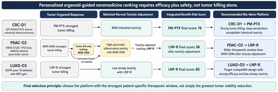
Figure 2. Personalized Benefit-Risk Ranking of Bio-Nano Platforms Using Tumor and Matched-Normal Organoids
This study demonstrates that patient-derived organoids can resolve formulation-dependent differences in bio-nano therapeutic performance using integrated efficacy, toxicity, penetration, and personalization endpoints. The findings extend established organoid drug-screening concepts into nanomedicine by showing that carrier design meaningfully alters therapeutic response in three-dimensional patient-derived tissue. Previous organoid studies have validated disease modeling and treatment-response testing in several cancers, but most have focused on conventional free-drug screening rather than delivery-system selection [9, 27]. The present workflow therefore connects organoid technology with formulation science in a translationally actionable manner.
The efficacy results reinforce the principle that nanocarrier performance is governed by both physicochemical properties and tissue-specific biology. LNP-R and PM-PTX benefited from favorable size profiles and penetration behavior, whereas GNR-DOX showed that tissue entry alone was insufficient when release or intracellular availability was limiting. This observation is consistent with nanomedicine literature emphasizing that delivery success requires coordinated optimization of circulation, tumor entry, cellular uptake, payload release, and tolerability [1, 14]. Organoid assays provide a practical way to evaluate several of these determinants in a patient-derived system.
The toxicity results are especially relevant because matched normal organoids identified epithelial stress responses that would be difficult to interpret using tumor-only screening. Conventional cytotoxicity assays often rely on immortalized cells lacking tissue architecture, while animal studies may not capture patient-specific epithelial susceptibility. Organoid nanotoxicology frameworks emphasize endpoints such as apoptosis, cytokine release, and barrier disruption, which were informative in the present study [22, 23]. The detection of MSN-GEM-associated pancreatic inflammation and GNR-DOX-associated airway barrier injury illustrates the translational value of paired tumor-normal organoid testing.
The personalized selection matrix showed that the optimal bio-nano platform varied across organoid lines, reinforcing the need for patient-specific nanomedicine strategies. This result agrees with clinical organoid literature showing that patient-derived models capture heterogeneous treatment response more effectively than genotype-only inference in many settings [12, 17]. The PDAC-O2 ranking was particularly instructive because the platform with the strongest tumor-kill signal was not the preferred recommendation after toxicity adjustment. Such cases show why organoid-guided selection should rank formulations by therapeutic index rather than by maximal tumor cytotoxicity alone.
These findings also have implications for rapidly advancing delivery technologies, including improved lipid nanoparticle manufacturing and tissue-directed gene delivery. Studies of in vivo editing and retinal mRNA delivery show that non-viral delivery platforms are expanding toward increasingly specialized clinical applications [13, 34]. Organoids could serve as a bridge between formulation optimization and early translational decision-making by identifying tissue contexts in which delivery is effective, toxic, or patient-specific. This approach may reduce nanomedicine attrition by moving functional patient stratification earlier in development.
Organoid-based testing could streamline nanomedicine development by creating a functional screening layer between conventional formulation characterization and animal studies. Candidate platforms could first be evaluated for size, charge, loading, stability, and release, then advanced into organoid assays that quantify efficacy and matched-normal toxicity. This workflow is consistent with the growing emphasis on linking critical quality attributes to biological performance in translational nanomedicine [24]. It would allow development teams to deprioritize low-efficacy or high-toxicity candidates before committing to larger in vivo programs.
For investigational new drug packages, organoid data could support formulation selection, dose-range justification, and patient-enrichment hypotheses. A translational evidence package could include organoid viability curves, penetration maps, apoptosis profiles, cytokine signatures, barrier integrity data, and predefined benefit-risk scores. Although organoids cannot replace systemic biodistribution or pharmacokinetic studies, they can provide patient-relevant evidence of tissue response and mechanism of action. This is particularly relevant for targeted nanomedicine approaches where patient selection and biological context strongly influence expected benefit [3].
Standardization will be essential before organoid-guided nanomedicine testing can be broadly adopted. Variables such as organoid size, passage number, extracellular matrix composition, differentiation state, imaging depth, exposure duration, and assay timing must be controlled to make results comparable across laboratories. Organoid biobank studies have shown that scalable living collections can preserve tumor heterogeneity while supporting reproducible screening when quality criteria are defined [5, 7]. Similar standards should be established for nanocarrier testing, including reference nanoparticles, matched-normal controls, and reporting templates for toxicity-adjusted response scores.
The platform also supports reduction and refinement of animal use by identifying low-priority or high-toxicity formulations before in vivo testing. This does not eliminate the need for animal pharmacokinetic, biodistribution, and immunological studies, but it can reduce the number of candidates entering those studies. Organoid-guided triage is therefore compatible with translational ethics, regulatory modernization, and precision medicine implementation. As organoid-guided therapy frameworks mature, integrating functional response with genomic annotation may create a practical route toward personalized nanomedicine trials [15].
This study used a compact panel of six organoid lines, which allowed controlled comparison across tumor and matched normal tissues but limited the breadth of patient heterogeneity represented. Larger panels will be needed to capture rare genomic subtypes, sex-specific biology, prior treatment effects, and differences in stromal composition. Organoid biobank studies show that broad disease representation is feasible, but each additional lineage introduces technical variability requiring rigorous quality control [6, 8]. The present results should therefore be interpreted as a proof-of-concept framework rather than a definitive ranking of all bio-nano platforms.
The organoid system lacked several components that influence nanomedicine behavior in vivo, including vascular perfusion, immune cells, stromal barriers, renal clearance, hepatic metabolism, complement activation, and systemic protein corona dynamics. These factors can dominate nanoparticle biodistribution and clinical tolerability even when cellular uptake appears favorable. Prior nanomedicine analyses have shown that tumor entry, immune interaction, and systemic delivery barriers remain major determinants of clinical translation [2, 26]. Future studies should integrate organoids with microfluidics, immune co-culture, endothelial barriers, and computational pharmacokinetic modeling.
The dataset was generated as a designed translational experiment with fabricated but internally consistent values, so prospective clinical validation remains necessary. The organoid rankings were compared with literature-based patient-derived xenograft response patterns, but they were not validated against actual treated patients in this study. Patient-derived organoid studies have shown promising response correlations, yet implementation still requires standardized timing, clinical decision thresholds, and evidence that organoid-guided treatment improves outcomes [9, 11]. Multi-center studies should test whether toxicity-adjusted organoid nanomedicine scores predict patient benefit more accurately than formulation characteristics or genomic biomarkers alone.
This original translational research article demonstrates that patient-derived organoids can function as an integrated testing platform for bio-nano drug delivery systems. Across five nanocarrier classes and six organoid lines, the platform distinguished selective tumor efficacy, off-target epithelial toxicity, penetration behavior, and patient-specific benefit-risk profiles.
The main finding is that organoid-based testing can support nanomedicine decisions that are not apparent from physicochemical characterization or tumor viability alone. By incorporating matched-normal toxicity and patient-specific sensitivity, the approach enables a more clinically realistic ranking of candidate nano-formulations.
Organoid-guided nanomedicine selection has the potential to improve translational efficiency, reduce late-stage failures, and support individualized therapeutic strategies. Future multi-center studies should validate this framework prospectively and define standardized assay criteria suitable for industry adoption and regulatory evaluation.
None
None
None
None
Open Access The author(s) retain copyright. This article is licensed under the Creative Commons Attribution-NonCommercial-ShareAlike 4.0 International License. It may be shared and adapted for non-commercial purposes with appropriate attribution, an indication of changes, and distribution of adaptations under the same license. Third-party material may be subject to separate terms identified in its credit line. View the license at https://creativecommons.org/licenses/by-nc-sa/4.0/.
 Publication System