Diabetes mellitus remains one of the most prevalent chronic diseases worldwide and is projected by the World Health Organization to become the seventh leading cause of mortality by 2030. In efforts to enhance therapeutic strategies for diabetes, mineral-based compounds are being explored for their supportive benefits. This study investigates the potential effects of the novel mineral formulation Dibeston on renal excretory function in a model of alloxan-induced diabetes. The experimental design involved 40 white laboratory rats assigned to five groups: Group 1: healthy controls; Group 2: alloxan-induced diabetes (untreated controls); Group 3: alloxan-induced diabetes treated with Dibeston; Group 4: alloxan-induced diabetes treated with Asparkam; and Group 5: alloxan-induced diabetes treated with a selenium-based preparation. Key renal parameters, including blood glucose, serum creatinine, urine output (polyuria), and urinary protein levels (proteinuria), were measured. The findings indicate that Dibeston significantly improves renal function in alloxan-induced diabetic nephropathy, suggesting its potential as an adjunct in diabetes management.
The global burden of diabetes mellitus continues to escalate, with current estimates from the International Diabetes Federation indicating approximately 450 million individuals affected worldwide—a figure that is expected to double every 12 to 15 years [1, 2]. Projections from the World Health Organization suggest that by 2030, diabetes mellitus will rank as the seventh leading cause of mortality [3]. Among its most serious complications is diabetic nephropathy, a progressive impairment of renal excretory function that contributes significantly to both disability and mortality in diabetic patients [4–6]. In particular, diabetic nephropathy occurs in nearly 45% of patients diagnosed with type 2 diabetes mellitus (T2DM), especially those who develop the condition during adolescence [7].
T2DM is commonly accompanied by a constellation of metabolic disturbances, including obesity, dyslipidemia, impaired glucose tolerance, hyperinsulinemia, hyperleptinemia, and blunted incretin responses. It also disrupts the endocrine balance, affecting thyroid and sex hormone levels [8–11]. These alterations stem from systemic metabolic dysregulation, heightened oxidative stress, chronic inflammation, and dysfunctional hypothalamic regulation of the endocrine axis [12, 13]. Central to this regulation are insulin and leptin, which cross the blood-brain barrier via receptor-mediated transport. However, persistent hyperglycemia and resistance to insulin and leptin compromise this transport, leading to deficiencies in hypothalamic signaling [14–17].
Reduced hypothalamic sensitivity to insulin and leptin impairs the synthesis and release of key releasing hormones such as thyrotropin-releasing hormone and gonadotropin-releasing hormone. These, in turn, regulate the secretion of thyroid-stimulating hormone and gonadotropins from the anterior pituitary [18–20]. Consequently, dysfunction in insulin and leptin signaling disrupts central appetite regulation and the systemic control of carbohydrate and lipid metabolism [21–23].
Given the complexity of T2DM and its complications, efforts to enhance treatment include developing novel therapeutic agents and supportive interventions. Mineral and vitamin complexes, antioxidants, and phytotherapeutic agents have shown promise as adjunctive therapies [24, 25]. This study aims to investigate the effects of a mineral complex containing magnesium, sodium, potassium, and selenium on blood glucose levels and renal excretory function in a rat model of alloxan-induced diabetes.
This study was conducted using adult male white Wistar rats, maintained under standardized laboratory conditions to ensure consistent nutrition, sleep-wake cycles, and overall care, in accordance with established guidelines for laboratory animals [26]. A total of 40 rats, each weighing 200-250 grams, were randomly assigned to five groups. The first group served as the intact control. The second group received an induction of diabetes mellitus using alloxan. The third group, also diabetic, was treated with Dibeston, a complex of mineral supplements. The fourth group, similarly diabetic, was administered the pharmaceutical preparation Asparkam. The fifth group consisted of diabetic rats treated with a selenium-based compound.
To induce diabetes, a freshly prepared 10% aqueous solution of alloxanhydrate was administered subcutaneously at a dose of 100 mg/kg body weight after an 18-hour fasting period. Dibeston was composed of sodium chloride (28%), potassium chloride (22%), potassium citrate (12%), potassium bromide (1%), magnesium sulfate (14%), calcium asparaginate (6%), magnesium asparaginate (8%), and glutamic acid (5%). This mixture was provided to the animals as a 1% solution available for unrestricted oral consumption.
Asparkam was administered as an oral solution in a dosage of 0.1 ml per 100 g of body weight. The solution, drawn from 10 ml ampoules, was delivered via gastric intubation for ten consecutive days before alloxan injection. The selenium preparation used, Selenopyran, was administered intramuscularly at a dose equivalent to 0.04 mg of selenium per 100 g of body weight, beginning three days prior to and continuing on the day following diabetes induction.
Post-induction, each treatment was administered for 10 additional days. The animals were then housed in metabolic cages for 24 hours to monitor urine output and water consumption. Following this period, the rats were euthanized for sample collection. Parameters evaluated included daily diuresis, fluid intake, urinary protein concentration, and glucose and creatinine levels in both blood and urine. Glomerular filtration rate and water reabsorption were calculated according to standard methodologies described in the literature [27–30]. Data were statistically analyzed using the Statistics software package.
The administration of Asparkam and Selenopyran resulted in a pronounced hypoglycemic effect, as evidenced by significantly reduced blood glucose levels—3.37 ± 0.30 µmol/L and 4.71 ± 0.35 µmol/L, respectively—compared to the hyperglycemic state observed in the untreated diabetic group (8.00 ± 0.22 µmol/L). In contrast, treatment with Dibeston did not yield a statistically significant reduction in glycemia (Figure 1).
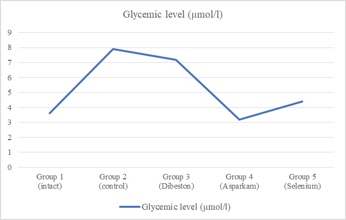
Figure 1. Blood glucose levels in rats following treatment with mineral supplements.
The induction of diabetes mellitus via alloxan was associated with marked renal dysfunction, including polyuria (7.34 ± 0.60 ml/100 g versus 3.20 ± 0.50 ml/100 g in control animals), excessive fluid intake (polydipsia), glucosuria, and proteinuria. Additionally, a decline in glomerular filtration and renal water reabsorption was noted, along with a significant elevation in blood creatinine concentrations (152.80 ± 10.30 µmol/L in diabetic rats versus 93.10 ± 2.20 µmol/L in intact controls). Treatment with the tested mineral agents ameliorated several of these renal abnormalities. Specifically, all three supplements reduced polyuria (Figure 2), with the most notable improvements observed in the Dibeston and Asparkam groups, where urine output was reduced to 2.00 ± 0.24 ml/100 g and 1.48 ml/100 g, respectively.
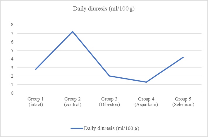
Figure 2. Polyuria levels in rats treated with mineral supplements.
Furthermore, treatment with the tested compounds diminished glucosuria and mitigated proteinuria. Notably, rats receiving Hyposol exhibited a substantial reduction in urinary protein excretion, with daily protein loss falling to 0.24 ± 0.02 mg/100 g compared to 0.75 ± 0.27 mg/100 g in untreated diabetic animals (Figure 3).
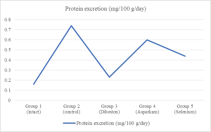
Figure 3. Proteinuria levels in rats following administration of mineral supplements.
In addition, Hyposol and Selenopyran were effective in lowering elevated serum creatinine levels. Treated animals exhibited creatinine concentrations of 82.0 ± 1.0 µmol/L and 110.2 ± 5.5 µmol/L, respectively, in contrast to 152.8 ± 9.3 µmol/L in the untreated diabetic group (Figure 4).
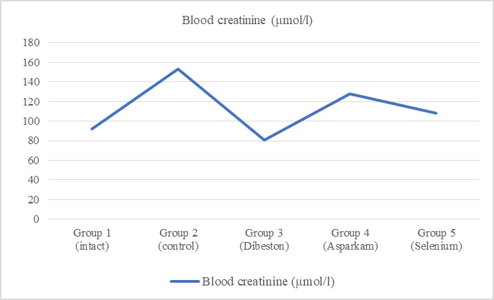
Figure 4. Serum creatinine concentrations in rats treated with mineral supplements.
These findings suggest that, in addition to their hypoglycemic effects (particularly with Asparkam and Selenopyran), the administered compounds exert a protective effect on renal function in alloxan-induced diabetes.
The observed improvements in renal excretory function following administration of the investigated compounds may, in part, be attributed to their hypoglycemic effects, as evidenced by reduced polyuria, polydipsia, and glucosuria, particularly in the Asparkam and Selenopyran treatment groups. In addition, Selenopyran’s pronounced antioxidant properties [31, 32] likely contribute to its renoprotective activity. The positive impact of Dibeston may be linked to its modulatory effect on the renin–angiotensin–aldosterone system, leading to reduced intraglomerular hypertension and improved renal hemodynamics [33].
This study provides evidence that mineral-based compounds, particularly Dibeston and Selenopyran, exert protective effects on renal excretory function in a rat model of alloxan-induced diabetes mellitus. These findings suggest that such compounds may offer therapeutic value in mitigating diabetic nephropathy and could be considered as potential components in the development of adjunctive treatments for diabetes, particularly in cases complicated by renal dysfunction.
None
None
None
None
Open Access The author(s) retain copyright. This article is licensed under the Creative Commons Attribution-NonCommercial-ShareAlike 4.0 International License. It may be shared and adapted for non-commercial purposes with appropriate attribution, an indication of changes, and distribution of adaptations under the same license. Third-party material may be subject to separate terms identified in its credit line. View the license at https://creativecommons.org/licenses/by-nc-sa/4.0/.
 Publication System