This article focuses on examining the structure of microorganisms and their antimicrobial drug resistance during the provision of in-hospital medical care for patients with COVID-19. Among monotypic fungal infections, Candida albicans predominated at 46.82% (n = 375), followed by Candida glabrata at 21.97% (n = 176). Streptococcus pneumoniae accounted for 25.24% (n = 617), while the combined antimicrobial presence of S. pneumoniae with Candida species represented 11.17% (n = 273), ranking highest in isolation frequency among all microorganisms. In deceased patients, the dominant pathogens identified were Klebsiella pneumoniae at 36.3% (n = 159) and Candida fungi at 21.92% (n = 96). Analysis of antimicrobial resistance revealed that S. pneumoniae (n=890) exhibited low resistance rates to benzylpenicillin (4.2%, n = 37), levofloxacin (11.5%, n = 102), and linezolid (0%), but showed elevated resistance to erythromycin (27.4%, n = 244), lincomycin (25.2%, n = 224), and doxycycline (15.5%, n = 141). The vast majority of K. pneumoniae isolates (n = 326) demonstrated resistance to ampicillin (97.9%), cefuroxime (94.2%), cefotaxime (87.7%), ceftazidime (85%), and cefepime (73%), with comparatively lower resistance to amoxicillin-clavulanic acid (52.1%, n = 170) and meropenem (33.4%, n = 109). Microorganisms exhibiting pan-drug resistance accounted for 7.6% (n = 125) of isolates, with the highest proportion of PDR strains attributed to K. pneumoniae, comprising 33.4% (n = 109) of all Klebsiella isolates. C. albicans and S. pneumoniae are the most frequently isolated pathogens among COVID-19 patients receiving inpatient care. The rising detection of extensively drug-resistant (XDR) and pan-drug-resistant (PDR) Klebsiella spp. underscores the critical need for careful selection of empirical antibacterial therapies.
Since late January 2020, new COVID-19 cases have been reported across numerous countries worldwide. On March 11, 2020, the World Health Organization (WHO) officially declared COVID-19 a pandemic [1, 2]. To date, it remains unclear whether the lung damage caused by COVID-19 in its classical presentation resembles the lung injury seen in infections such as SARS (caused by SARS-CoV), MERS (caused by MERS-CoV), or H1N1 pneumonia, which typically manifests as inflammation of the bronchopulmonary system of microbial and viral origin, often progressing to respiratory distress syndrome. Alternatively, it is questioned whether COVID-19 causes a distinct form of injury affecting not only the lungs but also organs like the kidneys, heart, and brain [3, 4].
According to A.G. Chuchalin, President of the Russian Respiratory Society and Academician of the Russian Academy of Sciences (RAS), the changes observed in lung tissue in COVID-19 patients are not typical pneumonia but rather pneumonitis characterized by diffuse alveolar damage and extensive fibrin deposition. This condition frequently leads to lung tissue fibrosis, which can be detected radiographically a month or more following disease onset. Fungal pathogens may contribute to triggering and sustaining this pathological process [5]. Professor Lev V. Kaktursky, Academic Advisor at the FSSI RI of Human Morphology and President of the Russian Society of Pathology, emphasizes the widespread involvement of not only the lungs but also other organs and tissues [6].
The most frequent complication of COVID-19 is acute respiratory distress syndrome (ARDS). Other documented complications include septic shock, acute kidney injury, myocardial damage, and secondary bacterial and fungal infections. Both non-invasive and invasive mechanical ventilation are employed to treat patients with these common complications; however, their use increases the risk of ventilator-associated pneumonia and other purulent-septic complications.
Pathogenetic therapy is widely used in COVID-19 treatment, involving glucocorticoids (dexamethasone, methylprednisolone), Janus kinase inhibitors (tofacitinib, baricitinib), IL-17 inhibitor (netakimab), IL-6 inhibitor (olokizumab), and IL-6 receptor blockers (tocilizumab, sarilumab, levilimab). A frequent adverse effect of these medications is an elevated risk of secondary bacterial infections [7]. Consequently, pathogenetic therapy in COVID-19 patients can increase the incidence and severity of secondary purulent-septic complications.
To address this, empirical antimicrobial therapy is necessary, guided by the microbial profile of causative agents involved in secondary purulent-septic complications and their antimicrobial drug (AMD) resistance patterns.
Furthermore, the occurrence of secondary bacterial infections in COVID-19 patients should be viewed in the context of nosocomial infections (NIs) or healthcare-associated infections (HAIs). Contemporary understanding of HAIs considers the dynamics of epidemic processes within medical organizations (MOs). A key criterion for classifying infections as HAIs is their direct link to medical care activities, including diagnosis, treatment, prevention, and rehabilitation. Thus, infections that develop not only as superinfections in hospitalized patients but also as a consequence of any medical interventions are classified as HAIs, including infections in healthcare providers themselves (Health and Safety Rules and Standards 2.1.3.2630-10).
Given the significance of pathogen spread within HAIs, addressing their regional and local circulation is critical, as is identifying effective antimicrobial drugs (AMDs). Additionally, managing infectious security strategies in medical institutions caring for COVID-19 patients presents considerable financial challenges.
The objective of this study is to investigate the structure of microorganisms and their resistance to antimicrobial drugs during the course of in-hospital medical care for patients diagnosed with COVID-19.
This research utilized bacteriological data obtained from biological samples, including sputum, bronchoalveolar lavage fluid, blood, and autopsy tissues, collected from COVID-19 patients treated at an infectious diseases hospital in 2020. A retrospective analysis was performed on 3,599 bacteriological test results from patients, along with 718 results from the Department of Pathological Anatomy.
Bacterial identification was conducted in accordance with regulatory guidelines governing the operations of bacteriological laboratories. The susceptibility of microorganisms to antimicrobial drugs was determined using the disk diffusion method. Interpretation of susceptibility levels followed the clinical guidelines outlined in the document “Identification of microorganisms’ sensibility to antimicrobial drugs” (as approved at the XVI International Congress of Antimicrobial Chemotherapy IACMAC/ESCMID, 2014).
Assessment of extreme drug resistance (XDR) and pan-drug resistance (PDR) among pathogens was carried out using internationally recognized clinical definitions, taking into account the specific identification of microorganism species.
Statistical analysis was conducted using descriptive statistics and the χ² test through the “MS Office EXCEL 2003” software. The significance threshold was set at P < 0.05.
Microbiological monitoring revealed that 67.94% of COVID-19 patients (n = 2,445) had positive bacteriological findings, indicating a notable role of microorganisms in the progression of the disease. Similar findings were recorded among deceased patients, with 61.0% (n = 438) showing resolutive bacteriological results. These findings suggest that approximately two-thirds of hospitalized COVID-19 patients developed secondary infections.
Among hospitalized COVID-19 patients, Candida spp. was isolated in 59.75% of cases (n = 1,461). Pure Candida spp. infections were identified in 32.76% (n = 801), while mixed infections involving Candida and other microorganisms accounted for 27.00% (n = 660). Among monotypic fungal infections, Candida albicans was most prevalent at 46.82% (n = 375), followed by C. glabrata at 21.97% (n = 176) (Table 1).
Table 1. Structure of results of Candida spp. selection from patients admitted to the hospital in terms of work at the infectious COVID-19 hospital in 2020
| Mono selection | Mixed infection with bacteria | Total | |||
| N | % | N | % | N | % |
C. albicans | 375 | 46,82 | 370 | 56,06 | 745 | 50,99 |
C. krusei | 129 | 16,10 | 130 | 19,70 | 259 | 17,73 |
C. tropicalis | 121 | 15,11 | 87 | 13,18 | 208 | 14,24 |
C. glabrata | 176 | 21,97 | 73 | 11,06 | 249 | 17,04 |
Candida spp. | 801 | 32,8 | 660 | 27,0 | 1461 | 59,75 |
Given the high isolation rate of fungi from the Candida spp., it is highly probable that these pathogens contribute to the pathogenesis of acute respiratory distress syndrome (ARDS) in patients with COVID-19 by sustaining the ongoing inflammatory response. This assumption highlights the need for further investigation into the pathomorphological alterations within this patient group.
Among the most commonly isolated microorganisms were Streptococcus pneumoniae (25.24%, n = 617), the microbial combination of S. pneumoniae with Candida spp. (11.17%, n = 273), and the association of Klebsiella pneumoniae with Candida spp. (8.3%, n = 203) (Figure 1). Collectively, pneumococcus was detected in over 36.4% of all positive cultures. In cases of mixed infection involving S. pneumoniae and Candida spp., the most frequent pairing was S. pneumoniae with C. albicans, representing 54.95% (n = 150) of such cases. In comparison, non-albicans strains (including C. Crusei and C. glabrata) comprised 30.4% (n = 83).
A comparable distribution was observed in mixed infections involving K. pneumoniae and Candida spp.. Here, K. pneumoniae combined with C. albicans was found in 58.13% (n = 118) of instances, whereas associations with non-Candida albicans strains accounted for 28.08% (n = 57).
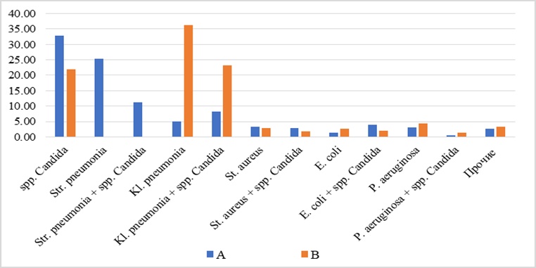
Figure 1. Structure of selected microorganisms received from patients admitted at the infectious COVID-19 hospital (n = 2445) (letter A) and from the Department of Pathological Anatomy (n = 438) (letter B) in 2020, %.
Among deceased patients, the dominant pathogens were K. pneumoniae at 36.3% (n = 159), the microbial combination of K. pneumoniae with Candida spp. at 23.29% (n = 102), and mono-isolation of Candida spp. fungi at 21.92% (n = 96) (Figure 1).
Within the K. pneumoniae/Candida spp. microbial association, non-albicans strains were most frequently isolated, with C. Crusei accounting for 56.86% (n = 58) and C. glabrata for 10.78% (n = 11) of all Candida isolates (Table 2). The composition of mono-isolated Candida spp. fungi in deceased patients showed statistically significant differences compared to those observed in hospitalized patients (P < 0.05). In this group, C. crusei was the most prevalent at 56.25% (n = 54), followed by C. glabrata at 13.54% (n = 13). Overall, fungal pathogens were detected in 50.46% (n = 221) of the deceased cases.
Table 2. Structure of results of Candida spp. selection from bacteriological research at the Department of Pathological Anatomy in terms of work at the infectious COVID-19 hospital in 2020, (%)
| Mono selection | Mixed infection with bacteria | Total | |||
| N | % | N | % | N | % |
C. krusei | 54 | 56,25 | 68 | 54,4 | 122 | 55,20 |
C. glabrata | 13 | 13,54 | 12 | 9,6 | 25 | 11,31 |
C. albicans | 14 | 14,58 | 28 | 22,4 | 42 | 19,00 |
C. tropicalis | 15 | 15,63 | 17 | 13,6 | 32 | 14,48 |
Candida spp. | 96 | 21,92 | 125 | 28,54 | 221 | 50,46 |
Based on the above findings, there are statistically significant differences in the microbial landscape between patients undergoing in-hospital care and those who are deceased. The microorganisms isolated from deceased patients predominantly belong to typical in-hospital pathogens, strongly suggesting the presence of healthcare-associated infections. The notably high contamination rate with Candida spp. is likely associated with ongoing pathogenetic therapy. At the same time, the frequent detection of K. pneumoniae correlates with the use of both invasive and non-invasive mechanical lung ventilation.
Invasive candidiasis, which typically occurs as a hospital-acquired infection in up to 12% of cases, is known to carry a high mortality rate of up to 70% [8]. On the other hand, bronchopulmonary candidiasis lacks specific clinical and radiographic features and can present as interstitial lung injury, which, on CT imaging, may resemble lung damage caused by COVID-19 [7, 9, 10]. Therefore, further investigation into the pathomorphological patterns of lung involvement in deceased COVID-19 patients is warranted.
The resistance analysis of leading microorganisms to antimicrobial drugs revealed that S. pneumoniae (n = 890) demonstrated low resistance to benzylpenicillin at 4.2% (n = 37), levofloxacin at 11.5% (n = 102), and linezolid at 0%. However, it showed high resistance to erythromycin (27.4%, n = 244), lincomycin (25.2%, n = 224), and doxycycline (15.5%, n = 141).
The proportion of oxacillin-resistant S. aureus (MRSA) strains was 38.9% (n = 58), and vancomycin-resistant S. aureus (VRSA) was found in 8.7% (n = 13). Notably, no linezolid-resistant staphylococcal strains were identified.
Among the isolates of K. pneumoniae (n = 326), a high level of resistance was observed: 97.9% to ampicillin, 94.2% to cefuroxime, 87.7% to cefotaxime, 85% to ceftazidime, and 73% to cefepime. Resistance to amoxicillin combined with clavulanic acid was relatively lower at 52.1% (n = 170), suggesting the production of extended-spectrum β-lactamases (ESBL) by this pathogen. Observations from clinical practice and patient monitoring trends indicate a growing resistance of gram-negative bacteria to carbapenems—a class once considered the last line of defense [9]. Alarmingly, 33.4% (n = 109) of K. pneumoniae isolates were resistant to meropenem, which is a concerning prognostic marker. A potential mechanism underlying resistance to β-lactams, including carbapenems, is the production of carbapenemases [11, 12]. Resistance to ciprofloxacin and amikacin was also notable, at 45.1% (n = 147) and 41.7% (n = 136), respectively.
Microorganisms exhibiting pan-resistance (PDR) constituted 7.6% (n = 125) of all isolates. K. pneumoniae accounted for the highest proportion among these, representing 33.4% (n = 109) of all Klebsiella isolates. Another concerning observation is the detection of PDR strains of E. coli, which made up 4.7% (n = 6). This may indicate plasmid- or transposon-mediated resistance mechanisms, which can facilitate rapid proliferation of PDR strains across the Enterobacteriaceae spp. group. Additionally, P. aeruginosa showed resistance to all classes of antimicrobial drugs in 10.23% (n = 9) of cases (Figure 2).
Regarding microorganisms with significant drug resistance (XDR–resistant to all but one or two classes of AMDs), the findings were as follows: 41.72% (n = 136) for K. pneumoniae, 10.24% (n = 13) for E. coli, and 18.18% (n = 16) for P. aeruginosa. These XDR strains act as a reservoir for potential future pan-resistant organisms.
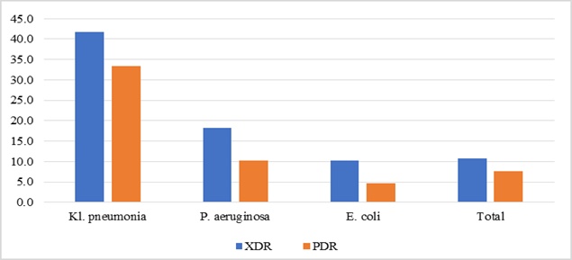
Figure 2. Selection of microorganisms with extensive drug resistance (XDR - microorganisms are resistant to all AMD but one or two classes) and pan-resistant microorganisms (PDR - resistant to all known classes of AMD) from clinical samples in 2020 (%).
The results obtained in this study underscore the importance of prescribing antimicrobial and antifungal drugs to hospitalized COVID-19 patients. Given the frequent detection of C. albicans and S. pneumoniae, agents such as amoxicillin and fluconazole may be considered appropriate treatment options.
Pathogens responsible for secondary infections in COVID-19 patients often display substantial resistance to antimicrobial drugs, including carbapenems. The growing incidence of K. pneumoniae strains exhibiting extreme or pan-drug resistance (PDR) underscores the urgent need for selecting effective empirical antibiotic regimens. In this context, special attention should be paid to the widespread presence of Klebsiella spp. as a common agent of healthcare-associated infections.
An informed selection of antimicrobial agents must rely on continuous microbiological monitoring to track the evolving resistance patterns in infectious disease hospitals. To limit the rise of antimicrobial resistance, it is vital to adopt a comprehensive strategy that includes evaluating the appropriateness of drug usage in COVID-19 management, along with monitoring the susceptibility of dominant pathogens to disinfectants.
1. A strong predictor of poor clinical outcomes in COVID-19 is the co-detection of PDR strains of C. krusei and C. glabrata.
2. All patients with ARDS in intensive care should be given initial antifungal therapy targeting C. krusei and C. glabrata, utilizing agents such as voriconazole, caspofungin, micafungin, or anidulafungin.
3. The high prevalence of K. pneumoniae with extreme and pan-resistance necessitates active surveillance of in-hospital strains and their resistance to disinfectants.
4. Considering the widespread use of immunosuppressive therapies in COVID-19 treatment and the increased risk of secondary infections, it is essential to routinely evaluate both the etiological spectrum of pathogens and the general profile of antibiotic resistance within infectious disease wards.
None
None
None
This research adheres to the ethical principles outlined in the Helsinki Declaration for medical research involving human subjects (2000 revision). Ethical approval was granted by the Ethics Committee of Far-Eastern State Medical University on June 21, 2019 (Protocol No. 5).
Open Access The author(s) retain copyright. This article is licensed under the Creative Commons Attribution-NonCommercial-ShareAlike 4.0 International License. It may be shared and adapted for non-commercial purposes with appropriate attribution, an indication of changes, and distribution of adaptations under the same license. Third-party material may be subject to separate terms identified in its credit line. View the license at https://creativecommons.org/licenses/by-nc-sa/4.0/.
 Publication System