Policosanol is a valuable compound with potential applications across multiple sectors. Within the pharmaceutical field, policosanol and its main components—triacontanol, octacosanol, and hexacosanol—have shown biological activity, particularly in conditions associated with inflammation and hypercholesterolaemia. Triacontanol, specifically, serves as a plant growth promoter and is widely applied in numerous economically significant crops and microalgae, either as a pure compound or as part of policosanol extracts. This review compiles key studies addressing the bioactivity of policosanol in both plant and animal cells, enabling comparison of the different mechanisms of action. A detailed evaluation of this information opens avenues for further research. Articles were sourced from PubMed and Redalyc using specific key terms: policosanol, inflammatory mechanisms, triacontanol, cellular absorption, photosynthesis, and photoinhibition. Policosanol has been found to interfere with inflammation-related pathways, notably the NF-κB and MAPK signaling cascades. Its cholesterol-lowering capacity results from the suppression of hepatic cholesterol synthesis through the indirect inhibition of HMG-CoA reductase. Triacontanol enhances plant growth and influences biochemical and physiological traits, especially under stress, mainly by improving photosynthetic efficiency. Notably, octacosanol can suppress the activity of triacontanol in plants—a phenomenon not observed in human cells—highlighting key distinctions in how these compounds function in plant versus animal systems, which warrants further investigation.
Policosanol (POL) refers to a complex blend of long-chain alcohols (LCAs), typically composed of 16 or more carbon atoms, characterized by a high molecular weight and the presence of a hydroxyl group. These compounds are solid, non-polar, and possess extremely high boiling points, while being notably insoluble in water at ambient temperature. POL derived from various waxy materials has demonstrated a range of advantageous effects on human health and has also shown potential as a growth promoter in both plants and cyanobacteria [1–3]. Although pharmacological evaluations have primarily focused on the complete mixture of LCAs within POL, individual constituents also exhibit distinct therapeutic benefits across different pathological conditions [4–6]. Compounds such as triacontanol (TRIA), octacosanol (OCTA), and hexacosanol (HEXA) have been extensively studied through in vitro and in vivo experiments [5, 6], with ongoing investigations into their specific mechanisms of action, metabolic pathways, and possible drug interactions [4, 5]. Moreover, formulation innovations, including the design of prodrugs, have been explored to enhance the delivery and efficacy of these lipophilic substances while minimizing bioavailability challenges [6, 7–9].
TRIA, in particular, is the only LCA identified to stimulate growth in plants and cyanobacteria [1]. Studies are ongoing to elucidate TRIA’s role as a plant growth promoter and how it interacts with other enhancers such as phytohormones and biofertilisers [10, 11].
In this context, this review aims to examine the action mechanisms of POL, highlighting both the shared and unique effects it exerts in animal and plant systems, to encourage further research into these biological processes.
This review compiles key findings from diverse studies that have documented the biological activity of policosanol and its constituent long-chain alcohols in both pharmacological and agronomic contexts. The literature search was conducted through PubMed and Redalyc databases, using the following key terms: policosanol, inflammatory mechanisms, triacontanol, cellular absorption, photosynthesis, and photoinhibition.
Numerous investigations have recognized the significant biological potential of policosanol (POL). While the mixture as a whole has shown beneficial effects, individual components also exhibit distinct pharmacological activities, contributing to the management of conditions such as hypertension, cancer, hyperlipidaemia, Parkinson’s disease, bacterial infections, gastrointestinal ulcers, and alopecia, among others [3].
Key LCAs, such as triacontanol (TRIA), octacosanol (OCTA), and hexacosanol (HEXA), have been subjected to various studies aimed at elucidating their mechanisms of action. Due to their structural similarity, it is hypothesized that these compounds may share comparable metabolic pathways [12]. For instance, investigations into OCTA have indicated that a portion of the compound undergoes catabolism and is transformed into fatty acids or chain-shortened derivatives via β-oxidation in peroxisomes [12, 13].
In terms of cellular absorption, earlier findings confirmed that enterocytes can take up POL, though comprehensive data on the exact internalization mechanisms remain limited. Given their structural resemblance and lipophilic properties, POL compounds are thought to enter cells through pathways similar to those used by fatty acids [13]. Fatty acid uptake by enterocytes typically occurs through two primary routes: passive diffusion and carrier-mediated transport [14]. Cocucci et al. [15] noted that the ability of lipophilic substances, including small therapeutic molecules and proteins, to pass directly through the plasma membrane is dependent mainly on their lipophilicity. Passive diffusion plays a significant role in drug uptake by enterocytes, whereas organs like the liver, kidneys, and brain rely more heavily on carrier-mediated systems [16–18]. Further research indicates that the absorption of very-long-chain fatty acids—and potentially long-chain alcohols—may depend on specialized protein-mediated mechanisms, such as those involving the scavenger receptor CD36, which is found on the surface of intestinal and hepatic cells [14].
To overcome the inherent limitations in the bioavailability of LCAs, specific strategies have been developed. For example, TRIA and OCTA can be conjugated with Polyethylene Glycol (PEG), significantly enhancing their solubility by up to 14 orders of magnitude. As an illustration, the solubility of unmodified TRIA is approximately 9 × 10⁻¹⁴ g/L, whereas PEGylated TRIA achieves solubility levels up to 10 g/L. Research has shown that these PEGylated nanoparticles are internalized by cells through macropinocytosis, a process facilitating polymer entry (Figure 1). This mechanism notably improves uptake by tumor cells and consequently enhances the anticancer potential of TRIA [6, 8].
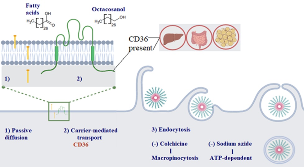
Figure 1. Schematic illustration of the mechanisms by which POL enters the cell: 1) POL may be internalized through passive diffusion, akin to fatty acids, owing to its comparable structure, low molecular weight, and lipophilic nature; 2) POL might also utilize fatty acids carrier-mediated transport via CD36, a transporter found in hepatocytes, enterocytes, and adipocytes; and 3) The cellular uptake of PEGylated TRIA occurs through ATP-dependent macropinocytosis.
Inflammation is a complex biological defense mechanism that arises from infections, physical trauma, or as part of specific disease processes. An increasing body of research indicates that POL and its associated compounds exhibit anti-inflammatory effects. The process is initiated when harmful stimuli, such as pathogens or tissue damage, are detected by receptors—referred to as sensors—located on immune cells like dendritic cells, mast cells, and macrophages. Additionally, inflammation is amplified by cytokines such as tumor necrosis factor-alpha (TNF-α) and interleukin-1 (IL-1), which stimulate the mitogen-activated protein kinase (MAPK) pathway. This activation leads to the stimulation of transcription factors, namely nuclear transcription factor-<kappa>B (NF-κB) and activating protein-1 (AP-1), within immune system cells. The MAPK pathway includes three central kinases: extracellular signal-regulated kinase (ERK), c-Jun NH2-terminal kinase (JNK), and p38. These enzymes initiate intracellular signaling cascades that control gene transcription related to inflammation, including the expression of cyclooxygenase (COX-2), inducible nitric oxide synthetase (iNOS), and various pro-inflammatory cytokines [19].
As immune cells become activated, they release several inflammation-mediating substances—such as cytokines, chemokines, nitric oxide (NO), prostaglandin E2 (PGE2), and leukotriene B4 (LB4). These mediators increase vascular dilation and permeability, allowing immune components, such as plasma, neutrophils, and leukocytes, to migrate into tissues, resulting in symptoms like edema, fever, and pain (Figure 2) [20, 21].
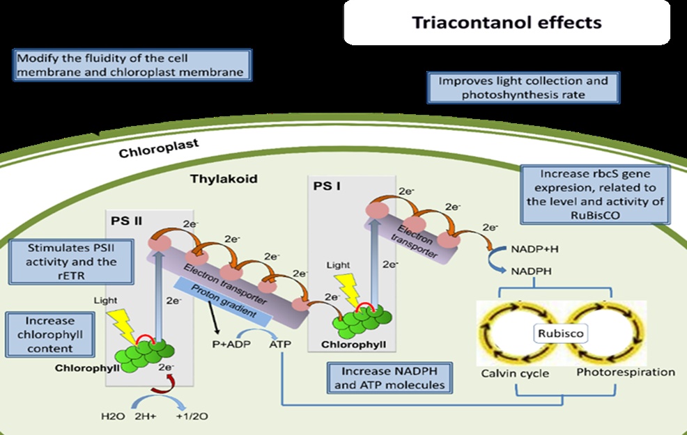
Figure 2. Effects of TRIA on photosynthetic activity. PSI: photosystem I; PSII: photosystem II; rETR: relative photosynthetic electron transport rate; ADP: adenosine diphosphate; ATP: adenosine triphosphate; NADP and NADPH: oxidized and reduced forms of nicotinamide adenine dinucleotide; RuBisCO: ribulose-1,5-bisphosphate carboxylase/oxygenase.
Research by Ravelo et al. [22] using acute inflammation models showed that D-002, a purified form of POL derived from beeswax, significantly suppressed oedema and reduced myeloperoxidase (MPO) enzyme activity. MPO, predominantly produced by neutrophils, contributes to host defense by generating reactive oxidants [23]. This reduction in MPO activity implies that D-002’s anti-inflammatory action may stem from decreased neutrophil accumulation in damaged tissues.
In another study, Fernández-Arche et al. [24] explored the nonglyceride fraction of pomace olive oil—containing HEXA, OCTA, and tetracosanol—and found it markedly inhibited the release of prostaglandin E2 (PGE2), nitrite, and TNF-𝛼 by 38%, 88%, and 83%, respectively, in macrophage models. This suppression was likely due to the downregulation of NF-κB activity, contributing to inflammation control [24]. In humans, the NF-κB pathway is responsible for upregulating iNOS and TNF-𝛼 and can be activated by lipopolysaccharide (LPS), oxidative stress, or cytokines [25]. Studies suggest that POL interferes with iNOS protein expression and helps moderate the excessive production of NO by inactivating NF-κB—a mechanism comparable to how statins function [24]. It is hypothesized that POL blocks NF-κB from entering the nucleus, although the precise molecular interactions remain to be fully determined.
Further supporting this mechanism, Guo et al. [4] found that OCTA reduced the nuclear presence of p65 and c-Jun—core subunits of NF-κB and AP-1, respectively. In addition, OCTA inhibited both transcription factors and suppressed the phosphorylation of p38 and JNK. This cascade of events led to reduced pro-inflammatory cytokine expression and a consequent decline in inflammation [4].
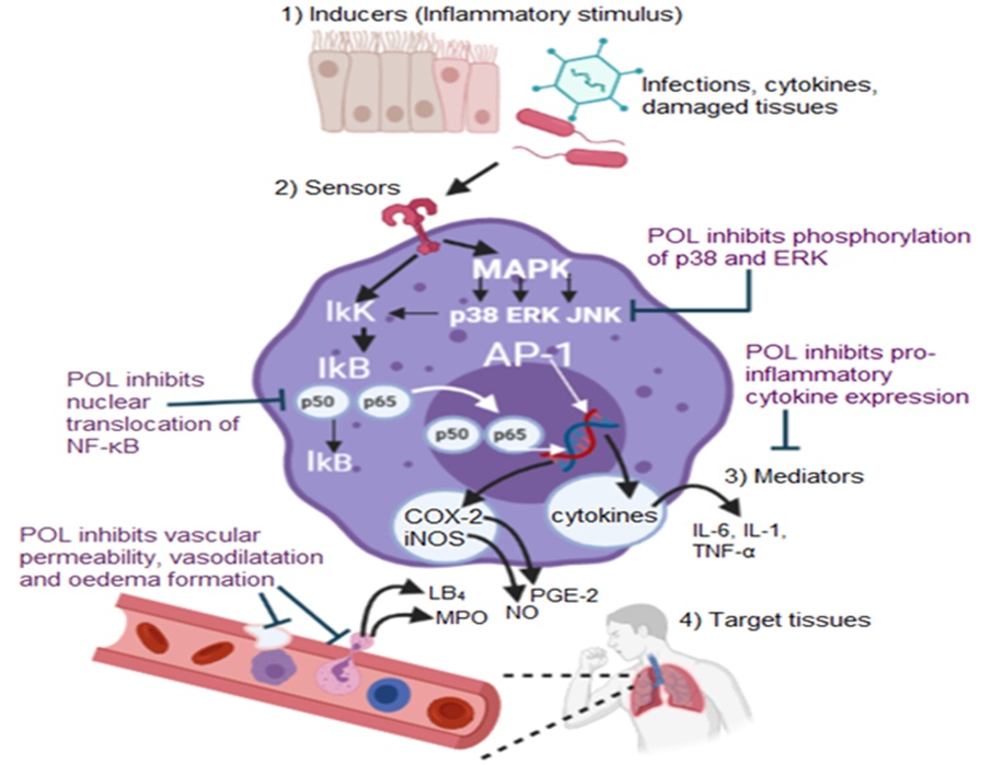
Figure 3. Molecular mechanisms underlying the anti-inflammatory action of POL. NF-κB refers to a group of dimeric transcription factors composed of five distinct protein monomers: p65, RelB, cRel, p50, and p52. Its activation is initiated when IκB inhibitor proteins are degraded, which permits the translocation of free NF-κB dimers—such as p50-p65—into the nucleus. Activating protein 1 (AP-1), another key transcription factor, is primarily composed of proteins from the c-Jun and c-Fos families and plays a crucial role in regulating inflammation-related gene expression.
In conclusion, POL, OCTA, and TRIA block the movement of NF-κB into the nucleus and inhibit MAPK phosphorylation, which together lead to a reduction in the production of inflammatory mediators. Additionally, POL reduces vascular permeability, oedema, and the enzymatic activity of MPO, decreasing neutrophil-derived reactive oxidants (Figure 3).
Inflammation is strongly connected to cardiovascular diseases and obesity, partly because maintaining cholesterol within normal ranges is crucial for healthy bodily function. In liver cells, cholesterol is synthesized internally via the mevalonate pathway, where acetyl-CoA is converted into cholesterol. A key reaction in this pathway is the transformation of 3-hydroxy-3-methyl-glutaryl-CoA (HMG-CoA) into mevalonate, a step catalyzed by the enzyme HMG-CoA reductase [26]. POL’s cholesterol-lowering properties are linked to multiple processes, especially the downregulation of HMG-CoA reductase through phosphorylation by AMPK (adenosine 5´-monophosphate-activated protein kinase), which inactivates the enzyme [5].
AMPK is a serine/threonine kinase composed of three subunits: a catalytic α-subunit, which can be phosphorylated at sites including Thr172, Thr258, and Ser485, along with regulatory β and γ subunits [26, 27]. AMPK orchestrates the balance between ATP generation and consumption, with its activation achieved through phosphorylation by various kinases and molecules. POL promotes phosphorylation of AMPK at Thr172 in hepatoma cells, thereby inactivating HMG-CoA reductase and lowering cholesterol synthesis [28]. Structurally, HMG-CoA reductase is a glycoprotein embedded in the endoplasmic reticulum membrane, where its catalytic carboxy-terminal domain faces the cytosol, and its amino-terminal domain anchors it within the membrane [29].
Nam et al. [30] showed in an animal model that POL decreases HMG-CoA reductase activity without altering its mRNA levels in hypercholesterolemic rats. This effect might result from increased AMPK phosphorylation triggered by an indirect pathway, though the detailed molecular mechanism was not investigated [30]. Subsequent work by Lee et al. [5] found that HEXA acts as an allosteric activator of AMPK by directly binding to its β subunit. They further demonstrated that HEXA phosphorylates HMG-CoA reductase at Ser872, which could reduce the enzyme’s affinity for NADPH [26]. These findings imply that other long-chain alcohols like OCTA or TRIA may share this AMPK activation mechanism. Furthermore, HEXA was observed to delay nuclear translocation of sterol-regulatory element-binding Protein-2 (SREBP-2), a key transcription factor controlling HMG-CoA reductase expression, suggesting that HEXA modulates SREBP-2 post-translationally by limiting its nuclear entry and thereby suppressing gene transcription [5].
Therefore, the mixture of POL or its individual LCAs (HEXA, OCTA, and TRIA) within liver cells—or their related fatty acids—may reduce cholesterol production through a dual mechanism: indirectly by promoting AMPK phosphorylation at Thr172 and directly by binding to the AMPK β-subunit, both leading to inhibition of HMG-CoA reductase activity [28].
Within the agricultural sector, TRIA stands out as the sole fatty alcohol explicitly utilized as a growth promoter for plants [1]. This contrasts with its effects on animal cells, where LCAs generally share similar modes of action. This difference suggests that plant cells uniquely recognize TRIA, enabling them to distinguish it from structurally similar molecules, such as OCTA, which differs by only two carbon atoms. Interestingly, OCTA can trigger distinct signaling pathways and may even inhibit the growth-promoting effects of TRIA [31].
Originally, TRIA was extracted from alfalfa (Medicago sativa) and later sourced from various plant and animal waxes. It has been tested across multiple species—including tomato, sweet pepper, sugar beet, cotton, tobacco, potato, and numerous fruit crops—with results demonstrating increases in dry and fresh biomass, plant height, as well as elongation of branches, stems, and roots. Furthermore, TRIA application has been linked to enhanced fruit and seed yields [1, 32, 33]. From a physiological perspective, TRIA promotes higher chlorophyll levels in leaves, improves photosynthetic rates, boosts nitrogen fixation, elevates enzymatic activity, enhances nutrient absorption, and stimulates the production of secondary metabolites and stomatal conductance [34]. Increases in essential nutrients such as nitrogen, phosphorus, and potassium have also been observed, alongside elevated levels of phenols, flavonoids, sugars, and proteins [35].
Beyond its field applications, TRIA has been employed in plant tissue culture techniques for micropropagation and for in vitro synthesis of secondary metabolites with medicinal value [1]. Additional benefits of TRIA include improvements in both quantity and quality of fruits, affecting traits such as length, width, firmness, Total Soluble Solids (TSS), acidity, and phenolic compound content [36].
TRIA’s growth-promoting properties extend beyond plants to cyanobacteria, where it enhances biomass production, photosynthesis, photorespiration, and chlorophyll and protein contents. These changes contribute to increased yields of fatty acid methyl esters (FAME), which hold significance for biodiesel manufacturing and pollutant removal via augmented cyanobacterial biomass [2].
One of the primary processes stimulated by externally applied TRIA is photosynthesis, with notable increases in parameters related to excitation energy capture efficiency by PSII reaction centers (Fv/Fm) [37], minimum fluorescence (F0), maximum fluorescence (Fm), electron transport efficiency of PSII, and photochemical quenching (qP), while concurrently reducing non-photochemical quenching (NPQ) [34, 37] and sustaining chlorophyll levels even under environmental stresses such as salinity, cold, and drought [1, 32, 35]. Kathuria et al. [38] reported that TRIA inhibits chlorophyllase activity but does not directly stimulate chlorophyll biosynthesis. Additional observed effects include modifications to chloroplast membrane fluidity and changes in the dynamic characteristics of protoplasts [39], increases in chloroplast number [34], and possibly improved thylakoid grana development.
Due to TRIA’s chemical structure—a 30-carbon aliphatic chain with low solubility—it cannot penetrate the plasma membrane, which serves as the initial barrier for plant growth promoters. Moreover, processes such as endocytosis and exocytosis are less active in plant cells compared to animal cells, where TRIA and other LCAs more readily enter through the membrane. Therefore, the specificity of TRIA’s action in plants may stem from the selective permeability of the plant cell membrane. It has been proposed that TRIA’s lipophilic properties alter membrane fluidity, as demonstrated by Ivanov and Angelov [39] in chloroplast protoplasts and supported by findings from Shripathi et al. [40], who studied microviscosity changes in cucumber fruit protoplast membranes (Cucumis sativus L.) [39, 40].
Membrane fluidity is influenced by both lipid composition and temperature. The hydrocarbon acyl chains of membrane phospholipids and glycolipids are primary factors in determining fluidity. However, this is also influenced by the size and charge of the polar head groups, as well as the amount of sterols present. The presence of short-chain or cis-unsaturated fatty acids tends to lower the membrane’s transition temperature, whereas saturated fatty acids and longer hydrocarbon chains raise it. Research by Swamy et al. [41] proposed that TRIA may act as a modulator by integrating into the inner membrane, thereby altering lipid phases and potentially activating membrane-bound enzymes. Shripathi et al. [40] highlighted differences between TRIA and abscisic acid (ABA), noting that ABA might be confined to specific membrane regions, unlike TRIA. These authors also observed that changes in membrane microviscosity could stem from variations in fatty acid composition as well as altered lipid-protein interactions, with both ABA and TRIA affecting the membrane’s physical state in distinct ways.
Phytohormones, or growth hormones, influence membrane fluidity due to their preferential affinity for the hydrophilic/hydrophobic interface of the membrane. The partitioning of these phytohormones and arbutin—a phenolic compound induced by drought—modifies the lipid phase, thereby affecting membrane properties such as permeability and microheterogeneity, which play key roles in cellular regulation [41]. At this initial stage, TRIA appears to modify how phytohormones partition within the membrane, triggering a signaling cascade that influences various metabolic processes. Swamy et al. demonstrated differential modulation by TRIA and jasmonic acid (JA) at the membrane level, which may explain TRIA’s antagonistic effects against JA [41]. Moreover, TRIA inhibits ABA’s activity [40], and since both ABA and JA are involved in stomatal closure during stress, TRIA’s role includes the reported inhibition of ABA-induced stomatal closure [34].
Regarding the distinct responses to TRIA versus OCTA, it has been reported that TRIA promotes the generation of a compound called TRIM at the tonoplast (the vacuole membrane). TRIM is believed to initiate a signaling cascade that enhances metabolic activity in plants. Conversely, OCTA, which antagonizes TRIA’s effect, induces the production of a second messenger termed OCTAM [31]. TRIM’s chemical structure is 9-β-L(+)-adenosine (9H-purin-6-amine, 9-β-L-ribofuranosyl) and is derived from adenosine, a component of Adenosine Triphosphate (ATP) and a precursor in cytokinin biosynthesis [42]. Therefore, the second messenger generated by TRIA (TRIM) may function similarly to cytokinins due to its structural resemblance, or it may provide adenosine or adenine for ADP and ATP synthesis, which are precursors in cytokinin formation. However, this action might also represent a modulatory response that either enhances or suppresses other phytohormones due to TRIA’s membrane-level effects. Supporting this, Kunjammal et al. [10] found a synergistic enhancement of growth and yield parameters in rice plants treated with a combination of TRIA (2 ppm) and cytokinin (10 ppm), compared to either treatment alone.
Beyond its role in hormonal regulation at the plasma membrane, TRIA also activates a second messenger (TRIM) and stimulates the enzyme NADP oxidase. Additionally, TRIA has been shown to increase ATP levels by activating ATPase [31]. The production of ATP is carried out by ATP synthetase, a multi-subunit complex that harnesses the proton motive force generated by the electron transport chain during photosynthesis. This proton gradient drives proton transport through ATP synthetase, facilitating the phosphorylation of ADP to ATP. This process predominantly takes place at the thylakoid membranes within chloroplasts, where ATP synthetase forms a critical component of the photosynthetic machinery responsible for energy synthesis.
When plants are exposed to light and heat stress, photosynthesis is inhibited due to oxidative damage targeting proteins of photosystem II (PSII). In particular, the D1 protein, which is integral to the PSII reaction center, is highly vulnerable to damage caused by reactive oxygen species (ROS) generated during photoinhibition. Heat-induced inactivation of PSII is often linked to lipid peroxidation occurring in proximity to PSII, which compromises the integrity of the D1 protein [43]. Furthermore, the fluidity of the thylakoid membrane is believed to be a key factor in maintaining PSII function during such stresses, as changes in membrane fluidity can disrupt the spatial organization of photosynthetic proteins and complexes, including ATP synthetase [43]. By modulating membrane characteristics, TRIA may influence ATP synthetase activity and photosynthetic efficiency, thereby regulating phytohormonal signaling either by enhancing or suppressing their actions and/or contributing to the synthesis of various compounds.
An elevation in photosynthetic activity leads to increased concentrations of ATP and NADPH molecules, both essential for the Calvin cycle and its regulation. The ATP present in the stroma can influence the light-induced activation of ribulose-1,5-bisphosphate carboxylase/oxygenase (RuBisCO) [44], while NADPH levels within the cell regulate the expression of genes involved in the Calvin cycle [45]. TRIA may enhance CO₂ fixation during the Calvin cycle by increasing ATP and NADPH levels, resulting in enhanced photosynthetic activity and upregulation of the rbcS gene, which is linked to increased RuBisCO quantity and activity [46]. Summarizing, TRIA’s function as a plant growth stimulator reported in various studies primarily targets photosynthetic enhancement (Figure 2); nevertheless, the primary mechanism behind these effects is not fully understood, leaving it uncertain whether the effects arise from the generation of a second messenger or mainly from alterations at the membrane level.
Separately, it is essential to highlight the protein SnRK1 (Snf1-related kinase1) in plants, which is homologous to the mammalian 5′ AMP-activated protein kinase (AMPK). Phosphorylation of SnRK1, triggered by POL, leads to the inhibition of HMG-CoA reductase and consequently reduces cholesterol biosynthesis. In plants, SnRK1 functions as a metabolic sensor that integrates a wide range of stress signals, playing a critical role in maintaining the energy balance necessary for growth and survival [47]. According to studies by Carianopol et al., SnRK1 may influence the abscisic acid (ABA) response by phosphorylating particular signaling molecules. The connection between SNF1 and AMPK is well established, given the conservation of regulatory processes such as T-loop phosphorylation, adenylate sensing, and kinase function; however, plants might regulate SNF1 through other, as yet unclear, mechanisms [48]. Therefore, uncovering the role of TRIA in SnRK1 activation could provide insights into how it enhances stress tolerance. SnRK1 and AMPK are pivotal in explaining the diverse biological effects seen in plants and animals, respectively. While AMPK activation is linked to numerous metabolic and therapeutic functions—such as anti-inflammatory and anticancer activities—SnRK1 participates in plant defense, primary and secondary metabolism, development, hormone responses to stress, and programmed cell death. Most of these processes have been reported to be influenced by TRIA, except programmed cell death in plants, which has not been investigated [47–49].
The diverse effects attributed to the long-chain alcohols (LCAs) comprising POL in both plants and animals prompt the question of whether their mechanisms of action share similarities. A notable difference lies in the selective response of plant cells to TRIA and the inhibitory action of OCTA, contrasting with animal cells, where responses to these LCAs tend to be more alike. This discrepancy may be partly explained by the lower levels of exchange and endocytosis observed in plant cells compared to animal cells.
Regarding TRIA’s mechanisms in plants, research remains limited and mainly descriptive, focusing on its ability to enhance fruit yield and quality, increase secondary metabolite production, and stimulate various physiological and morphological traits. Available studies propose that TRIA acts initially at the membrane level, triggering the formation of a second messenger. Some authors suggest that this response bears considerable resemblance to the action of cytokinins, phytohormones extensively utilized to promote multiple physiological processes and crop yields, and which may also play a role in delaying plant senescence. Nonetheless, further research is necessary to clarify this mechanism and to compare TRIA’s membrane-level effects with other physiological responses.
Lastly, the presence of SnRK1 proteins in plants, which function analogously to AMPK in maintaining cellular homeostasis, along with their similar activation mechanisms as orthologous proteins, indicates a potentially shared pathway between plant and animal cells. This could explain the range of effects observed in both cell types, with differences arising from membrane specificity, internalization, and distinct signaling pathways.
We express our gratitude to Consejo Nacional de Ciencia y Tecnología (CONACYT) and the Instituto Politécnico Nacional (IPN) for providing scholarships.
None
None
None
Open Access The author(s) retain copyright. This article is licensed under the Creative Commons Attribution-NonCommercial-ShareAlike 4.0 International License. It may be shared and adapted for non-commercial purposes with appropriate attribution, an indication of changes, and distribution of adaptations under the same license. Third-party material may be subject to separate terms identified in its credit line. View the license at https://creativecommons.org/licenses/by-nc-sa/4.0/.
 Publication System