Snakebite envenoming remains a significant global health challenge, with high mortality and morbidity rates persisting despite decades of medical attention. Each year, millions are affected by venomous snakebites, often resulting in death or severe disability. Snake venoms exhibit diverse bioactivities, including hemorrhagic, inflammatory, cytotoxic, cardiotoxic, and neurotoxic effects, mainly due to complex mixtures of toxin-rich proteins. Although considerable research has been undertaken, the majority of venom components remain uncharacterized. Recent advancements in proteomics and bioinformatics have enabled more detailed exploration of venom profiles, facilitating the identification and functional prediction of novel toxins. Computational approaches now enable modeling of toxin-target interactions, aiding understanding of venom mechanisms. This review also explores the emerging role of medicinal plants in snakebite treatment, alongside conventional antivenoms. Emphasis is placed on the urgent need to improve access to safe, affordable, and effective antivenoms in low-income tropical regions and to promote their appropriate clinical use.
Snakebite envenoming is a neglected tropical disease that continues to affect vulnerable populations in many regions [1]. It occurs when a venomous snake inadvertently injects venom into a human through specialized fangs connected to venom glands [2, 3]. The complex and variable composition of snake venoms results in a wide array of biochemical activities and toxic effects, leading to diverse clinical manifestations [4]. While some venom components cause localized tissue destruction with potential long-term consequences, others can trigger systemic effects such as neurotoxicity, resulting in respiratory muscle paralysis, acute kidney injury, rhabdomyolysis, cardiotoxicity, nervous system disturbances, and thrombosis [5–7].
Members of the Viperidae family typically produce venom that induces both local and systemic effects, including hemorrhage, coagulation disorders, and hypovolemic shock [8]. In contrast, venom from snakes of the Elapidae family primarily causes neurotoxicity, such as neuromuscular paralysis [9]. The Colubroidea superfamily, also known as advanced snakes, includes over 2,500 species with a wide geographical distribution and long evolutionary history [10]. All venomous snake species are classified under the Caenophidia clade, within the order Squamata and suborder Serpentes [11]. Some of the deadliest snakes belong to the Viperidae (true vipers and pit vipers) and Elapidae (including cobras, kraits, mambas, and sea snakes) families [12, 13].
Because snakes are ectothermic, they thrive in warmer climates, making tropical regions—especially in Latin America, Africa, and Oceania—most susceptible to snakebite incidents [14]. In these areas, human-snake encounters are more likely during the rainy season, which coincides with both peak agricultural activity and snake mating periods. Epidemiological data from hospital records underscore the significant burden of snakebite envenoming in terms of mortality and long-term disability [15]. However, community-based surveys in some countries suggest that actual mortality figures may far exceed those reported by healthcare facilities. In contrast, residents of high-income countries, such as those in North America and Europe, face a substantially lower risk of snakebites and are generally unaware of the global health crisis that snakebite envenoming represents [16].
This lack of awareness has contributed to a historical neglect by funding bodies, pharmaceutical companies, public health institutions, and advocacy organizations, limiting progress in addressing the social and medical challenges posed by snakebites. Despite being a substantial source of morbidity and mortality in tropical regions, snakebite envenoming remains underrecognized [17]. Cardiovascular complications are particularly underreported, primarily due to the scarcity of reliable data and the fact that most bites occur in remote, underserved areas [18].
This review evaluates the cardiovascular effects of snakebite envenoming and proposes a clinical algorithm for screening for cardiac involvement. Following a systematic review, several studies documenting cardiovascular complications were analyzed. Although relatively rare, such complications—when present—can lead to severe conditions, including myocarditis, myocardial infarction, ventricular dysfunction, hypotension, and cardiac arrest [19]. In fact, cardiovascular symptoms accounted for severe adverse outcomes in 24.39% of the reviewed cases, including fatal arrhythmias and cardiac failure [20]. Therefore, patients with systemic symptoms should undergo close clinical monitoring, comprehensive physical examination, and early electrocardiographic (ECG) evaluation to detect potential cardiac involvement. A schematic overview of the review structure is presented in Figure 1.
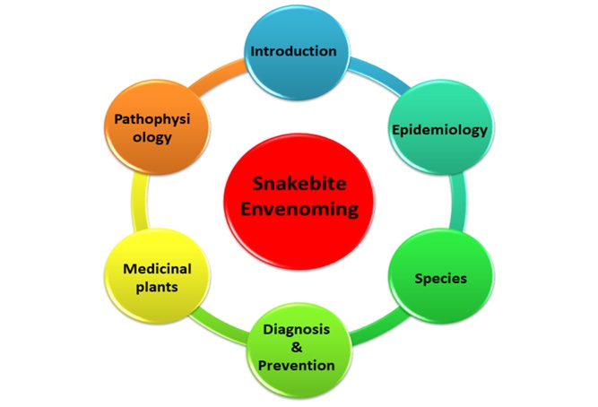
Figure 1. Graphical representation of Snakebite Envenoming.
This review also explores the emerging role of medicinal plants in snakebite treatment, alongside conventional antivenoms.
Snakebite envenoming poses a significant public health burden in many developing regions, particularly across tropical and subtropical zones. Areas most affected include Sub-Saharan Africa, South and Southeast Asia, Latin America, and Papua New Guinea. Globally, it is estimated that 1.8 to 2.7 million individuals suffer snakebite envenoming each year, resulting in approximately 81,410 to 137,880 deaths annually (Figure 2) [21]. India alone accounts for an estimated 46,000 of these fatalities [22]. In Sub-Saharan Africa, annual mortality is estimated to range between 7,000 and 30,000, although these figures are likely underreported due to limited data availability [23].
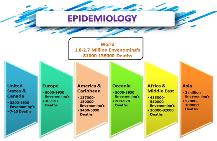
Figure 2. A global overview of snake envenoming and deaths.
Snakebites disproportionately impact economically disadvantaged populations, particularly those living in poor housing conditions and lacking access to healthcare, education, and services. Countries with low Human Development Index (HDI), low gross domestic product (GDP), and limited health expenditure face the greatest burden. The financial strain of treatment, forced borrowing, and loss of income often pushes affected families further into poverty. The most affected demographic is individuals aged 10-40 years—typically the most economically productive members of rural communities [24, 25]. Notably, children under five exhibit a higher case fatality rate. In India, for instance, the highest mortality rate due to snakebite envenoming was observed in children aged 5–14 years. Children are particularly vulnerable when engaging in outdoor activities such as farming, playing, or reaching into rodent burrows [26].
Snakebite envenoming is predominantly occupational and environmental in origin, with young people and agricultural workers among the most at risk. The specific populations affected vary by region. For example, sugarcane workers in South Africa, Saint Lucia, and Martinique; tea pickers in southern India; and rice paddy farmers in Myanmar—all face heightened risk. In Myanmar, snakebite envenoming ranks as the fifth leading cause of death and primarily affects agricultural laborers. Fishing communities in warm tropical waters are also vulnerable, particularly those using hand traps or ropes, and exposure extends to fishermen working in farming environments [27]. Pregnant women are especially at risk, with snakebites linked to fetal loss, maternal mortality, abortions, and antepartum hemorrhages in countries such as Nigeria and Sri Lanka [28].
Moreover, envenoming is a major environmental hazard among indigenous and nomadic populations, hunter-gatherers, and impoverished rural dwellers. This includes groups such as the Hadza in Tanzania, the San (Bushmen) of Southern Africa, and the Fulani and Turkana pastoralists in West Africa and Kenya. Similar vulnerabilities are observed among tribal and marginalized groups in India, Sri Lanka, and South America [29]. In regions such as the Amazon rainforest and the coastal plains of New Guinea, snakebite envenoming contributes significantly to the high mortality seen among native populations, including the Yanomami and Waorani ethnic groups.
Snakebite envenoming can result in long-term physical, psychological, and social consequences. Chronic outcomes include limb amputations, post-traumatic stress disorder (PTSD), myopia, muscle contractures, malignant ulcers, persistent infections, fetal loss, and maternal mortality. In Sub-Saharan Africa alone, an estimated 6,000 amputations occur each year directly due to snakebite, excluding cases from other African regions [30]. Even when excluding the burden of chronic disability, premature mortality caused by snakebite in India accounts for approximately 2.97 million disability-adjusted life years (DALYs). Globally, the estimated burden rises to 6.07 million DALYs [31]. These figures surpass the mortality associated with venomous animal encounters reported in the 2013 Global Burden of Disease study, highlighting a persistent issue of underreporting in snakebite data [32].
In West Africa, the combined burden of snakebite envenoming—including premature death and long-term disability—exceeds that of several other neglected tropical diseases (NTDs), such as Buruli ulcer, echinococcosis, leprosy, trachoma, yaws, yellow fever, and podoconiosis, across at least 16 countries [33]. Both early death and disability contribute significantly to the total impact of snakebites [34]. Nevertheless, in Sub-Saharan Africa, diseases such as trypanosomiasis, leishmaniasis, and onchocerciasis still collectively impose a higher burden than snakebites [35].
Venoms have evolved independently in diverse animal taxa, including snakes, spiders, scorpions, and jellyfish, serving both predatory and defensive functions. In snakes, venom acts as a vital trophic adaptation, essential for hunting and prey immobilization [36]. Its evolution reflects intense Darwinian selection, leading to the rapid diversification of venom proteins. These proteins, often working alone or in combination, are specialized to incapacitate prey or inadvertently harm human victims [37, 38]. Across taxa, venom traits are shaped by varying degrees of genetic complexity, with changes in toxin-coding genes and their expression likely contributing to venom diversity. However, the evolutionary mechanisms behind this vast biological variety remain only partially understood [39].
The evolution of animal venoms is analogous to other evolutionary innovations, such as eyes, wings, or fins—outcomes of convergent evolution in which similar traits emerge independently in distinct lineages. Neofunctionalization, wherein duplicated genes acquire novel functions, plays a pivotal role. These genes often encode structural motifs suited for toxic activity, forming complex venom compositions through gene duplications and multimeric protein interactions [40, 41]. Only a few multigenic protein families dominate venom profiles, yet these families exhibit substantial intra- and interspecific variability (Figure 3).
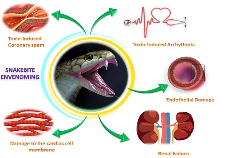
Figure 3. Toxic effects of snake venom toxins on the human body. Venoms have a wide range of toxic effects on the human body, and their composition determines which are most harmful. Neuromuscular paralysis caused significant tissue damage to the victim, and increased permeability of the blood vessels caused by viperid venoms, and cardiovascular shock might occur.
Understanding the ecological and evolutionary drivers that have shaped venom composition is crucial for delineating the phylogenetic and biogeographic distinctions among snake species. Mapping venom variability across spatial and temporal scales can reveal how local adaptations influence venom profiles. Insights into these processes can bridge the fields of evolutionary biology and clinical toxicology, informing the development of more targeted and effective antivenom therapies. Particularly, the most clinically relevant venom components in humans are often those that evolved to cause maximal incapacitation in natural prey. A deeper understanding of the adaptive mechanisms governing venom evolution will thus aid in the creation of next-generation snakebite treatments with enhanced specificity and efficacy [42, 43].
The growing scientific interest in venom biology has driven the advancement of “omics” approaches, which focus on the qualitative and quantitative profiling of venom constituents. Among these, venom proteomics has emerged as a pivotal technique. The integration of next-generation transcriptomics and proteomics technologies now enables exceptionally detailed characterization of snake venoms. This has led to an in-depth understanding of the relative abundance and diversity of major toxin families in the venoms of both viperid and elapid snakes [44, 45].
A direct translational application of venom proteomics is the study of antivenom immune responses—an approach known as antivenomics. This methodology quantifies the degree of immunological cross-reactivity between antivenoms and various homologous or heterologous venoms. Antivenomics, when combined with in vivo neutralization assays, provides a robust framework for assessing the potential clinical efficacy of antivenoms even before human trials commence. This dual approach enhances the preclinical evaluation of antivenom products and informs their therapeutic deployment [46].
Snakebite incidents, similar to other medical emergencies, require rapid and accurate diagnosis due to the potential for life-threatening complications. However, uncertainties about the snake species involved often complicate clinical decision-making, the amount of venom injected, and the venom’s composition, which can vary with the snake’s age, species, and geographic distribution [47].
In many rural and remote areas, snakebite victims are initially managed by registered nurses, community health assistants, or local medical personnel in primary care settings such as clinics and health centers [48]. Depending on the severity of the envenomation, patients may be referred to tertiary hospitals equipped with intensive care units and specialized diagnostic laboratories for further management.
When envenomation is suspected, immediate and thorough clinical assessment is critical to guide life-saving interventions. A detailed patient history should be obtained to document the onset and progression of symptoms. Rapid clinical evaluation should include assessment of vital signs to detect hypovolemia, postural changes in heart rate, signs of neurotoxicity such as ptosis and respiratory paralysis, and evidence of systemic bleeding.
Notably, bites from kraits (Bungarus spp., family Elapidae) often occur during sleep, with victims waking in the night to find a bite. Differentiating snakebites from other causes of puncture wounds or envenomation—such as arthropod bites (e.g., spiders), lizard or rodent bites, or plant thorn injuries—is essential for accurate diagnosis [19, 49]. Some bites may be“dry bites” where a venomous or non-venomous snake inflicts a bite without injecting venom, resulting in minimal or no symptoms.
If available, identification of the snake species—either through a photo or physical specimen—by a trained herpetologist can assist in guiding treatment [38, 50]. Witnesses and patients themselves can also provide valuable diagnostic clues by describing the bite event and recognizing clinical syndromes associated with specific types of envenomation.
Laboratory evaluations play a vital role in confirming systemic envenomation and guiding clinical management following snakebites. One of the early hematological indicators is peripheral neutrophilic leukocytosis, signifying an acute inflammatory response. Hematological findings may vary: anemia (low hematocrit) suggests hemorrhagic complications, whereas hemoconcentration (elevated hematocrit) points to plasma extravasation due to increased capillary permeability [51].
Evidence of microangiopathic hemolytic anemia, marked by the presence of schistocytes on a peripheral blood smear, is often associated with acute kidney injury, especially when accompanied by severe thrombocytopenia and a bleeding tendency. Systemic envenomation, including that caused by viperids, sea snakes, and certain rear-fanged colubrids, often results in coagulopathies. A key bedside diagnostic test—the 20-minute whole blood clotting test (WBCT20)—can help determine whether the blood remains incoagulable at room temperature, indicating anticoagulant venom exposure and a potentially life-threatening condition [52].
Further laboratory investigations to assess coagulopathy include prothrombin time (PT), activated clotting time (ACT), fibrin degradation products, and D-dimer levels to identify disseminated intravascular coagulation (DIC). Extremely elevated serum creatine kinase (CK), often above 10,000 U/L, indicates substantial skeletal muscle damage. Renal function should be monitored closely in all patients showing signs of acute kidney injury, including serum potassium evaluation and urinalysis for proteinuria and hematuria upon admission.
Cardiac involvement may manifest as subtle ECG abnormalities such as atrioventricular block, ST–T wave changes, bradycardia, or even myocardial ischemia. Individuals with pre-existing cardiovascular conditions are especially susceptible to myocardial infarction, often precipitated by hypotensive shock. Advanced imaging techniques like echocardiography may detect complications such as pericardial effusion, cardiac failure, or internal hemorrhages in the pleural or peritoneal cavities [31]. Wound ultrasonography is increasingly used to assess localized tissue damage, while CT or MRI is useful for identifying intracranial bleeding or infarction.
The retrospective identification of the offending species, evaluation of prognosis, and assessment of antivenom effectiveness can be facilitated through enzyme immunoassays (EIA), which detect and quantify venom antigens in various body fluids. Commercial venom detection kits capable of delivering results within 15–30 minutes are currently available only in Australia (manufactured by Seqirus) [53]. Although these kits are highly sensitive, they may lack the specificity required to distinguish between venoms of closely related species, and the mere detection of venom at a wound site does not necessarily indicate the need for antivenom therapy.
Biological samples suitable for venom antigen testing include tissue from the bite site, incision fluids, blister aspirates, serum, and urine. These specimens are also valuable for forensic investigations [54]. Other molecular approaches, such as reverse transcription polymerase chain reaction (RT-PCR) and the identification of snake DNA from bite-wound swabs, enable detection of venom-gland mRNA, providing species-level confirmation of the envenoming agent.
Preventive strategies against snakebites should prioritize community-centered education programs targeted at high-risk populations [55]. Dissemination of information through local media—including radio, television, mobile applications, social media, posters, street performances, and public meetings—can effectively raise awareness. Educational campaigns should emphasize behavioral modifications such as safe walking, working, and sleeping practices to reduce snakebite risk [56].
In regions where formal ambulance services are lacking, alternative transport solutions—such as boats or motorbike ambulances operated by trained community volunteers—can expedite access to medical care. Public health messaging must also discourage the use of traditional healers, as these interventions are generally ineffective and may delay access to appropriate treatment.
Immediate first aid is essential following a snakebite and should be administered by the victim or nearby individuals. Key components include reassuring the patient, immobilizing the affected limb, and removing constrictive items such as rings or tight clothing. Application of pressure immobilization techniques—such as pressure pads or elastic bandages over the bite site—can reduce venom dissemination through the lymphatic and venous systems. Rapid, yet calm, transportation to the nearest medical center is crucial. Analgesia should be limited to paracetamol (acetaminophen) or opioids; nonsteroidal anti-inflammatory drugs (NSAIDs) and aspirin should be avoided due to their potential to exacerbate bleeding [57].
To reduce the risk of fatal respiratory obstruction from bulbar paralysis or aspiration, the patient should be placed in the recovery position during transfer, and an oropharyngeal airway may be inserted to maintain upper airway patency. Harmful interventions such as incisions at the bite site, suctioning, and tight tourniquets must be strictly avoided. For suspected neurotoxic envenoming—particularly from species such as Acanthophis and certain Micrurus spp.—first-aid administration of atropine (an antimuscarinic) and neostigmine (an acetylcholinesterase inhibitor) has been proposed, given their potential actions at postsynaptic neuromuscular junctions [58].
All individuals reporting snakebite exposure should undergo at least 24 hours of clinical observation. Before removing any compression bandages or tourniquets, it is imperative to establish intravenous access and have resuscitative measures readily available. If the patient presents with signs of respiratory failure—such as cyanosis of the lips, tongue, or mucous membranes—airway management and supplemental oxygen should be initiated immediately. Intravenous fluids should be promptly administered in cases of circulatory shock. Pain severity can vary, and when present, it should be managed in accordance with established analgesic protocols [59].
Notably, patients may appear stable initially but can deteriorate rapidly within hours. Published envenoming severity scores are often unreliable as they are based on artificial or non-validated criteria. Enzyme immunoassays used to quantify venom antigen levels in plasma have been shown to assist in prognosis estimation. Continuous monitoring is essential to detect clinical features of envenoming, such as ptosis, spontaneous hemorrhage, or progressive local swelling. Urinary output should be closely monitored to assess renal function. In cases where compartment syndrome is suspected—due to massive muscular swelling confined within fascial compartments—intracompartmental pressure should be evaluated.
Neurotoxic envenoming poses a diagnostic challenge due to flaccid paralysis, which can obscure the assessment of consciousness with conventional tools such as the Glasgow Coma Scale. Patients may be unable to speak, move, or respond; however, if cardiorespiratory support is sufficient, communication can still be achieved through voluntary movements, such as toe or finger flexion, in response to yes/no questions when eyelids are manually lifted.
The decision to administer antivenom is the most critical therapeutic judgment following initial resuscitation and suspected species identification. Antivenom remains the only specific pharmacological agent capable of reversing the systemic effects of snake envenoming [60, 61]. Antivenoms are derived from large domestic animals—typically horses, sheep, or camels—that have undergone prolonged immunization with one or more snake venoms. To minimize hypersensitivity reactions, the immunoglobulin G (IgG) molecules are frequently enzymatically cleaved using pepsin to remove the Fc portion, yielding F(ab’)₂ fragments. Some manufacturers use papain digestion to generate smaller Fab fragments, enhancing tissue penetration but potentially increasing the risk of recurrent envenoming. Whole IgG preparations, often purified via caprylic acid precipitation, are still widely employed in the formulation of several antivenoms [62].
Affinity chromatography is increasingly used to purify antivenom immunoglobulins, thereby enhancing the safety and economic feasibility of production. Antivenoms targeting the most clinically significant snake species within a specific geographic region are referred to as polyvalent (polyspecific) antivenoms. For instance, Indian polyvalent antivenoms are formulated to neutralize venoms from the“big four” species: Naja naja and Bungarus caeruleus (family Elapidae), as well as Daboia russelii and Echis carinatus (family Viperidae). In contrast, monovalent (monospecific) antivenoms are directed against a single species’ venom, such as ViperaTab (Flynn Pharma), which is specific for Vipera berus (family Viperidae). Antivenoms have remained the cornerstone of treatment for over a century, demonstrating efficacy in mitigating many of the life-threatening and tissue-destructive consequences of envenoming [63].
When administered promptly, antivenoms can neutralize the effects of venom-induced coagulopathy, hypotension, and postsynaptic neurotoxicity, and may also attenuate or prevent presynaptic neurotoxicity, rhabdomyolysis, and local necrosis. Thus, determining whether to administer antivenom constitutes the most critical therapeutic decision in managing snakebite envenoming [64]. The selection of an appropriate antivenom depends on accurately identifying the offending snake species. Due to their highly specific neutralizing capability, antivenoms are only effective against venoms used in their immunization process or those of closely related species.
Antivenoms pose logistical challenges: they can be costly, are often scarce in high-need regions, and typically require cold-chain storage for transport and preservation. The cessation of production by major manufacturers such as Syntax, Behringwerke, and Sanofi Pasteur—primarily due to economic constraints—has led to severe shortages of antivenom, particularly in African nations.
Only a subset of snakebite incidents necessitates antivenom administration. Both adult and pediatric patients receive identical doses. Ideally, the initial dosage should be informed by human clinical trial data; however, in their absence, dosing is usually based on the manufacturer’s extrapolated neutralizing potency derived from rodent ED₅₀ studies. If symptoms such as progressive neurotoxicity or cardiovascular instability persist 1–2 hours post-administration, or if coagulation remains impaired at 6 hours, repeat dosing is warranted. Intravenous infusion is the standard route of administration, with infusion times ranging from 10 to 60 minutes [65].
Continuous monitoring is essential during the first 2 hours of antivenom administration, as this is the window when allergic and febrile reactions most commonly occur. Prophylactic intramuscular adrenaline administration has been shown to reduce both the frequency and severity of these early reactions. Should hypersensitivity reactions arise—manifesting as pruritus, urticaria, nausea, agitation, tachycardia, or tachypnea—prompt treatment with intramuscular adrenaline is essential.
In addition to antivenom, supportive treatment must address the systemic and organ-specific consequences of envenoming. In patients with bulbar dysfunction and respiratory paralysis, airway management via supraglottic or endotracheal intubation—either manual or mechanical ventilation—is critical [66]. Acetylcholinesterase inhibitors such as neostigmine, when co-administered with atropine, may temporarily improve neuromuscular function but do not substitute for antivenom therapy. The “ice pack test,” which evaluates bilateral ptosis improvement following localized cooling, may predict responsiveness to acetylcholinesterase inhibitors, such as the short-acting agent edrophonium.
Persistent hypotension or shock unresponsive to antivenom therapy necessitates cautious volume resuscitation and the use of vasopressors such as dopamine. If conservative measures fail to reverse acute kidney injury, renal replacement therapy must be initiated. A tetanus toxoid booster is universally recommended following a snakebite. However, prophylactic antibiotic therapy is not advised unless the wound is necrotic, secondarily infected, incised, or presents with abscess formation [66, 67]. In such cases, broad-spectrum antibiotics are warranted. Surgical intervention, including debridement or skin grafting, may be required in severe cases of tissue necrosis, and in extreme cases, amputation of gangrenous digits or limbs may be necessary.
Compartment syndrome—commonly affecting the anterior tibial compartment—is a recognized complication of envenomation. Although fasciotomy (a surgical procedure to relieve pressure in fascial compartments) is sometimes considered, it is infrequently indicated. In most cases, intracompartmental pressure remains within normal limits. Performing a fasciotomy before effective correction of venom-induced coagulopathy can result in catastrophic bleeding. Therefore, unnecessary surgical intervention may increase both hospitalization time and long-term morbidity [68].
Polyherbal combination therapies are gaining attention for their cost-effectiveness and therapeutic potential. While not yet widely implemented in clinical practice, this approach merits thorough investigation due to its numerous advantages, including affordability, reduced risk of adverse reactions, lack of expiration concerns, room-temperature stability, local availability, ease of administration, and potential to serve as both a primary and adjunctive treatment in cases of snakebite, particularly where antivenom serum (AVS) is unavailable. However, like all therapeutic modalities, polyherbal remedies are not without limitations, including low yield, variability due to geographic origin, and potential toxicity [69, 70]. Thus, it is imperative to conserve biological diversity and elucidate the mechanisms by which these plant-derived compounds exert their antagonistic effects on snake venom.
This review underscores the rich and diverse ethnomedical knowledge held by communities worldwide concerning medicinal plants. In contrast, the availability and reliability of conventional antivenoms remain problematic due to their high cost, limited accessibility, and risk of eliciting hypersensitivity reactions in susceptible individuals. Moreover, further investigation is warranted to determine whether the protective effects of plant-derived compounds—such as hepatoprotective, anti-inflammatory, antioxidant, and anticancer activities—extend to mitigating the systemic effects of snake venom [71]. Continued exploration and validation of new plant-based therapies are essential to advance the field of complementary and alternative medicine. Furthermore, refinement of polyherbal formulations, whether in crude or purified form, is necessary. A multidisciplinary research approach is crucial for integrating these therapies into broader treatment strategies.
The present study aims to document the use of traditional medicinal plants employed by African healers in the management of snakebite envenomation, thereby contributing to the preservation and dissemination of this indigenous knowledge. Numerous knowledge gaps exist regarding the clinical efficacy and safety of these botanical therapies, underscoring the need for rigorous scientific validation. Various medicinal plants have demonstrated promising antivenom activity in preclinical studies, highlighting the potential of herbal medicine as a supplementary or alternative approach [72, 73].
Ultimately, the primary objective of any healthcare system—whether traditional or modern—is to prevent and alleviate human and animal suffering. Accordingly, the following recommendations are proposed to support this goal.
Comprehensive scientific investigation is essential to evaluate the traditional herbal remedies employed by indigenous healers for the treatment of snakebites [74]. Findings from such studies should inform evidence-based guidelines and protocols for traditional healers, with a strong emphasis on dosage, safety, and efficacy. Furthermore, traditional practitioners must be held accountable for the responsible use of herbal therapies. Empirical evidence indicates that certain medicinal plants possess demonstrable efficacy against snakebite envenomation [75]. Therefore, a concerted effort must be made to collect, document, and analyze these plant resources.
Phytochemical and pharmacological studies will be critical in identifying bioactive compounds and validating the therapeutic potential of these plants. Instead of dismissing traditional medicine as outdated or ineffective, governmental and institutional support should focus on fostering the most successful aspects of indigenous healing systems. Providing incentives and institutional support for traditional healers will not only legitimize their practices but also encourage intergenerational transmission of valuable ethnomedical knowledge [76].
Table 1 shows that snake bites were traditionally treated using medicinal plants.
Table 1. Snake bites were traditionally treated using medicinal plants
Plant | Family | Local name | Part of the plant was used | References |
Cyphostemma junceum | Vitaceae | Etse Zewe | Chewing roots | |
Gossypium herbaceum L. | Malvaceae | Tit | Chewing root | |
Pergularia daemia L. | Asclepiadaceae | Yeayit Hareg | Root | |
Plumbago zeylanicum L. | Plumbaginaceae | Amira | Chewing leaves | |
Combretum molle G. Don | Combretaceae | Muama, Kiama | Root | |
Conyza sumatrensis | Asteraceae | Yadh asere, yadh | A leaf infusion of the plant | |
Entada leptostachya | Fabaceae | Mwaitha | Crushed stem | |
Opilia amentacea Roxb. | Opiliaceae | Mutonga | Roots crushed powder | |
Solanum incanum L. | Solanaceae | Mutongu | Stem and fruit dried powder | |
Microglossa pyrifolia | Asteraceae | Nyabungodidi, | Chewing leaves and juice | |
Boscia angustifolia | Capparidaceae | Kermed | Root stem bark | |
Nicotiana tabacum | Solanaceae | Timbhako | Leaves | |
Solanum incanum | Solanaceae | Engulle/sengol | Root |
Recent advancements in proteomics have led to a rapid expansion of knowledge about the peptides and proteins in animal venoms, particularly snake venom [80]. These developments have greatly improved the ability to detect a wide range of toxins in snake venom. This achievement was previously challenging with older techniques such as reversed-phase high-performance liquid chromatography (RP-HPLC) and mass spectrometry. However, with the vast amounts of data generated by modern “omic” methods, effective computational approaches are essential for analyzing and extracting meaningful information. The number of toxins identified using these advanced methods is substantial, and this review will emphasize databases that facilitate the search and retrieval of information related to snake toxins [50, 81]. Furthermore, this article will discuss computational tools developed to investigate snake venom activity, focusing on the prediction of toxin three-dimensional structures and their interactions with molecular targets. We will evaluate the advantages and limitations of molecular modeling for predicting binding affinities and specificities, and the future implications of integrating these computational insights into snake venom research.
Bioinformatics tools and databases, such as UniProt, NCBI GenBank/GenPept, and the Protein Data Bank (PDB), provide easy access to protein sequences and three-dimensional structures [82-84]. However, these resources lack standardization, especially regarding toxin nomenclature and their pharmacological activities, complicating efforts to mine data for snake venom peptides. Additionally, the reliance on author-submitted data often results in duplicated entries, and some venomous animal data remains unpublished in general databases. Emerging specialized venom databases, such as Conoserver for cone snail venoms, Arachnoserver for arachnid venoms, and ISOB (Indigenous Snake Species of Bangladesh), are helping fill this gap [85]. The Swiss Institute of Bioinformatics (SIB) has also introduced Venom Zone, a resource that compiles venom data from six species, including snakes. This database organizes its content by taxonomy, activity, and venom protein families, making it easier for users to access relevant information. Moreover, Venom Zone links its data to well-established protein databases, such as UniProtKB/Swiss-Prot and UniProtKB/Trembl, which are manually reviewed and annotated, thereby further enhancing the quality of the available information [86, 87].
The study of well-characterized peptides can provide valuable insights into the functionality of uncharacterized peptides and proteins. Specialized databases play a critical role in accessing this data, predicting the three-dimensional structures and functions of toxins, and identifying potentially novel toxins with unique properties. However, due to the lack of a standardized method for annotating toxic substances, estimating the total number of toxins in a particular animal’s venom remains challenging. One promising solution to this issue is the use of machine learning-based classifiers. Tools like Tox Classifier can distinguish toxin sequences from non-toxin sequences, enabling accurate classification of toxins into appropriate families and significantly enhancing the quality and utility of these databases [19, 88].
Snake venoms are complex proteomes composed of various peptides and proteins. A recent review has outlined the proteomic methodologies applied to investigate these components [89]. Techniques commonly employed in this context include electrophoresis, liquid chromatography, Edman degradation, amino acid analysis, enzymatic digestion, and mass spectrometry. Among these, high-performance liquid chromatography (HPLC), Edman degradation sequencing, MALDI-TOF/MS, one- and two-dimensional PAGE, and ESI/MS/MS of digested proteins are among the most widely used approaches.
The proteomic study of snake venom begins with venom extraction, a process referred to as “milking,” in which a snake is induced to bite into a suitable receptacle to release its venom. Following collection, protein separation is achieved using methods such as reverse-phase HPLC (RP-HPLC), ultra-high-performance liquid chromatography (UHPLC), and ion-exchange chromatography [90]. The resulting protein fractions are then analyzed for peptide sequence determination using mass spectrometry and Edman degradation. Before tandem mass spectrometry (MS/MS), peptides are typically reduced, alkylated, and enzymatically digested. The tertiary structures of these biomolecules are further examined via nuclear magnetic resonance (NMR) spectroscopy [91].
One of the defining characteristics of snake venom proteins and peptides is the prevalence of cysteine residues that form stabilizing disulfide bridges. These covalent linkages are critical for maintaining structural integrity, enhancing proteolytic resistance, refining selectivity, and supporting functional activity. Notably, the number of disulfide bonds can differ among venom peptides, contributing to their functional diversity [92].
Effective management of snakebite envenoming demands a globally coordinated response, guided by the WHO’s multi-tiered strategy and involving a broad coalition of stakeholders. Realizing such an initiative will require significant time, funding, and collaboration from UN Member States, global health agencies, researchers, and antivenom producers. Bioinformatics has emerged as a vital tool in elucidating the pharmacologically active constituents of snake venoms—from gene and protein identification to modeling three-dimensional structures and target interactions. This review highlights how such computational strategies have deepened our understanding of venom biology.
Nonetheless, predicting the specificity of snake venom toxins remains a major challenge due to their complex interactions with diverse ion channel subtypes, each of which elicits distinct physiological effects. Compared to traditional drug discovery models that focus on single targets, phenotypic screening may offer a more promising avenue for identifying novel therapeutic compounds. In this context, bioinformatics-led prescreening has the potential to uncover new molecular entities that diverge from known venom profiles, paving the way for innovative treatments.
None
None
None
None
Open Access The author(s) retain copyright. This article is licensed under the Creative Commons Attribution-NonCommercial-ShareAlike 4.0 International License. It may be shared and adapted for non-commercial purposes with appropriate attribution, an indication of changes, and distribution of adaptations under the same license. Third-party material may be subject to separate terms identified in its credit line. View the license at https://creativecommons.org/licenses/by-nc-sa/4.0/.
 Publication System