Zinc is a crucial element for both plant and human growth, playing a key role in cell division and supporting the maintenance of normal cellular processes. This study investigates the effects of zinc on mitotic division at various concentrations and time points. By assessing the correlation between the mitotic index (MI), chromosomal aberrations (IAC), and exposure time to different concentrations of zinc sulfate (ZnSO4), the cytotoxic effects of ZnSO4 were evaluated. The results showed a strong positive correlation (r > 0.89) between the incidence of chromosomal aberrations and higher concentrations of ZnSO4, suggesting that increased zinc levels contribute to chromosomal damage. Furthermore, increasing exposure time from 24 to 72 hours and concentrations from 10 ppm to 50 ppm resulted in a significant negative correlation (r = -0.84). These results highlight the critical role of zinc in cellular function and the potential harm from excessive zinc.
Zinc is an essential trace metal widely present in the human body and plays a critical role in various biological functions [1, 2]. Research has highlighted zinc’s diverse bioactive effects, including its roles as an antioxidant, anti-inflammatory agent, anticancer compound, and immunomodulator [2-5]. Zinc is also a key element for plant growth, as it requires a proper nutrient balance to ensure healthy development and optimal crop production. In humans, a significant portion of zinc intake comes from plant-based foods, especially in the form of ZnSO4 [6, 7].
This metal is integral to the structure of numerous proteins involved in key cellular processes such as RNA transcription, and it affects RNA breakdown, decreases RNA polymerase activity, alters ribosomal structures, and reduces ribosome numbers [4, 8, 9]. Zinc is unique as it is necessary for the proper functioning of all six enzyme classes—oxidoreductases, transferases, hydrolases, lyases, isomerases, and ligases [2, 10-12].
In plants, soil zinc availability is influenced by several factors, including concentration, ion speciation, and interactions with other essential nutrients, particularly ZnSO4 [2, 11, 13]. Zinc deficiency in crops leads to significant reductions in both yield and quality [14, 15]. Furthermore, insufficient ZnSO4 in the soil impacts the nutritional quality of crops by lowering the zinc content in their edible parts [3, 6, 13]. Typically, visible signs of zinc deficiency in plants only appear in severe deficiency [7, 16, 17].
Different plant genotypes exhibit varying degrees of tolerance to low ZnSO4 levels, impacting both zinc uptake and its use within the plant [18, 19]. While the molecular and physiological mechanisms underlying this tolerance are still under investigation, these insights could be leveraged for crop breeding to improve nutrient efficiency [16, 20-22]. Molecular markers associated with micronutrient efficiency (including ZnSO4 and Mn) have been identified in major crops like barley, wheat, and maize [1, 23-25].
Inadequate ZnSO4 levels in the soil hinder critical physiological functions in plants, negatively affecting their growth and development [26, 27]. ZnSO4 deficiency is considered a major threat to global food security [3, 7]. Micronutrient deficiencies, including ZnSO4, affect about 40% of the worldwide population, with ZnSO4 deficiency affecting approximately 2 billion people, according to the World Health Organization [28, 29]. This deficiency is most commonly seen in populations that rely heavily on cereal-based diets with low zinc content [3, 7]. Zinc accumulation in the brain has been linked to cell death and toxicity [3, 10, 30, 31]. This research aims to evaluate the genotoxic and cytotoxic effects of various ZnSO4 concentrations over a range of time periods (24–72 hours) using the Allium test.
The study used garlic bulbs (Allium sativum L.) from the Cenad-Timiș, Romania population, all of equal size, for the experimental procedure. The four experimental conditions included: V1 = control water (-H₂O), V2 = ZnSO4 at 10 ppm, V3 = ZnSO4 at 20 ppm, and V4 = ZnSO4 at 50 ppm, to assess their effects on growth and development.
Garlic bulbs were directly exposed to solutions corresponding to the four experimental treatments, and growth was monitored at 24, 48, and 72 hours. The focus was on examining the mitotic activity within the meristematic tissue of the roots.
At 24, 48, and 72 hours, the root tips of the bulbs were collected from each treatment group. These samples underwent a series of preparatory steps, including prefixing, fixation, hydrogenation, and staining, to preserve the biological material for analysis.
To assess the cytotoxic and genotoxic effects of ZnSO4 at the cellular level, the mitotic index (MI%) and the chromosomal aberration index (IAC%) were calculated using the Allium test [32-34].
Root tip sampling occurred in the morning to capture the period of maximal mitotic division activity. The collected root samples were fixed in Carnoy’s solution for 24 hours. Following fixation, the roots were hydrolyzed in hydrochloric acid (HCl) for 6 minutes in a 60 °C water bath. Subsequently, they were stained using Carr’s reagent. The prepared slides were analyzed using an Optika microscope [35, 36].
Cytological evaluations focused on cells at various mitotic stages (prophase, metaphase, anaphase, and telophase), as well as on chromosomal anomalies, including anaphase bridges, multipolar anaphase, isolated chromosomes, incorrect chromosome polarization, polyploidy, and binucleate cells.
To calculate the results, specific equations were employed as outlined in previous studies [35, 37].
The influence of zinc concentrations on mitotic activity in the root apex varied with exposure times (24, 48, and 72 hours). Elevated zinc levels induced toxicity, resulting in chromosomal defects that hindered normal mitotic progression and diminished root growth.
The mitotic index, calculated using Eq. (1), varied with treatment duration and ZnSO4 concentration (Figure 1). At the lowest concentration (V2- 10 ppm), mitotic activity remained comparable to the control (V1-H₂O) within the 24-48 hour window. However, at higher concentrations (twenty ppm and fifty ppm), a significant decrease in mitotic activity was observed during the 24-48 hour period, indicating a dose-dependent reduction in cellular activity.
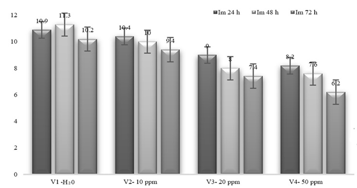
Figure 1. Mitotic index observed in Allium sativum species at various ZnSO₄ concentrations.
Zinc ions disrupted the normal progression of the mitotic phases, with the extent of disruption varying with both treatment concentration and exposure duration. Various concentrations of ZnSO₄ induced a range of chromosomal abnormalities across nearly all stages of mitosis, though the occurrence rates differed, as illustrated in Figure 2. The chromosomal aberration index (IAC) increased with increasing ZnSO₄ concentration, as shown in Figure 2. The highest percentage of chromosomal irregularities was observed at the highest ZnSO₄ concentration, specifically in the V4 experimental variant during the 24h-72h time frame.
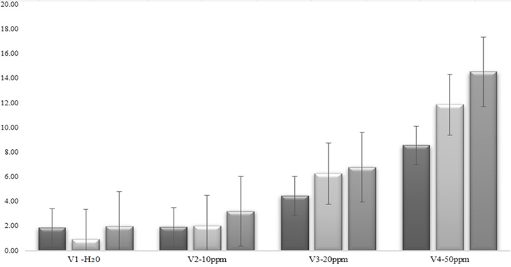
Figure 2. Index of chromosomal abnormalities in Allium sativum L. at varying concentrations of ZnSO₄.
As the chromosomal aberration index (IAC) was calculated, a noticeable increase in mitotic abnormalities was observed at both higher ZnSO₄ concentrations and longer exposure durations. Intermediate levels of chromosomal aberrations were observed in the V3 group during the 24–72 hour exposure period. Prolonged exposure (72h) to ZnSO₄ resulted in a significant rise in IAC across all treated variants. Even at the lowest V2 concentration (10 ppm), with a shorter exposure time (24-48h), the chromosomal aberration index (IAC) was similar to that in the control (V1-H₂O).
Graph a in Figure 3 shows a strong positive correlation between the number of dividing cells and the mitotic index, with an r-value of 0.987. This supports the expectation that as the number of cells increases, the mitotic index also rises. However, when analyzing the relationship between exposure time and the mitotic index, a weak negative linear correlation (r = -0.40, graph b) was observed during the 24–72 hour window. This indicates that time negatively influences the mitotic index (MI). The highest mitotic index was observed at 24 hours, while the lowest was at 72 hours. Additionally, a strong negative correlation (r = -0.84, graph c) was found between the concentration of ZnSO₄ and the number of cells in division. Lastly, the data shown in graph e of Figure 3 suggests that ZnSO₄ has a detrimental effect on both the number of cells undergoing division and the mitotic index.
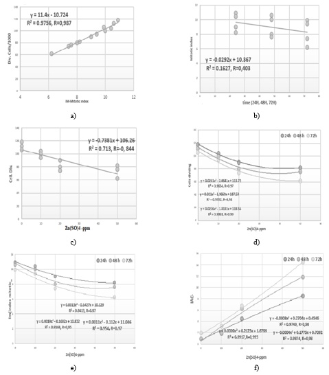
Figure 3. The outcomes of the post-experimental correlation analyses are illustrated as follows: a) relationship patterns observed between the population of cells undergoing division and the mitotic index, b) association trends between the mitotic index values and temporal progression, c) correlation dynamics identified between the number of dividing cells and ZnSO4 exposure, d) combined correlation profiles involving dividing cell counts, ZnSO4 treatment, and elapsed time, e) interaction correlations assessed among mitotic index, ZnSO4 concentration, and time factors, and f) correlation outcomes highlighting the interplay between index of chromosomal aberrations, ZnSO4 administration, and the time variable.
An inverse correlation was observed between zinc concentration and the number of dividing cells—higher zinc doses were associated with fewer dividing cells. In the control group (V1, no ZnSO4), cell counts remained between 100 and 120. In contrast, the V4 group treated with 50 ppm ZnSO4 exhibited the lowest cell division rates, ranging from 60 to 80 cells. Referring to graph e in Figure 3, it is evident that increasing ZnSO4 levels exerts a suppressive effect on cell proliferation. The untreated control variant (V1-H2O) consistently displayed the highest frequency of dividing cells (100–129), whereas the V4 treatment (ZnSO4-50 ppm) showed the greatest decline (approximately 60–80 cells). Moreover, division activity was greatest at the 24-hour time point across all groups, while it declined markedly by 72 hours, especially at the highest ZnSO4 concentration of 50 ppm.
As shown in graph f of Figure 3, ZnSO4 exposure also corresponded with a reduction in the mitotic index. A clear negative trend was evident: mitotic index declined with increasing zinc concentration. The control group (V1), which received no ZnSO4, showed the highest mitotic index (9.9%-10.9%), whereas treatment with ZnSO4 at 50 ppm (V4) resulted in significantly lower indices (6.0%-8.9%). The decrease in the mitotic index was especially notable when ZnSO4 levels rose from 20 ppm (V3) to 50 ppm (V4), particularly at longer exposure durations of 24 to 72 hours.
These observations align with previous findings reported in the literature, where zinc’s cytotoxic properties were shown to be dose-dependent. Specifically, higher concentrations of zinc were associated with an increase in abnormal mitotic figures, whereas lower concentrations had minimal to no adverse effects on the mitotic index [36, 38, 39]. Figure 4 presents microscopic images of the various mitotic stages in the plant cell.
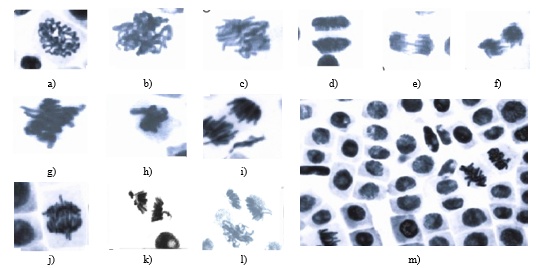
Figure 4. Microscopic depictions of mitotic progression within plant cells are shown, including (a and b) typical prophase, (c) standard prometaphase, and (d) conventional telophase, captured following exposure to varying levels of ZnSO₄; images (e and f) illustrate distorted anaphase with evident chromosomal bridges observed at 50 ppm, while (g) depicts prematurely aligned chromosomes during metaphase at 20 ppm. Further, panels (h and i) present metaphase and anaphase chromosomes appearing prematurely aligned at 20 ppm; in (j), anaphase is marked by the presence of spherical chromosomal configurations alongside precociously progressing chromosomes at 50 ppm; (k and l) exhibit anaphase figures with accelerated chromosomal movements, and (m) provides an overall microscopic field overview.
The analysis of the chromosomal aberration index (IAC%) reveals a pronounced, directly proportional association with both zinc concentration and exposure duration, ranging from 24 to 72 hours. A strong positive correlation (r > 0.9) was identified, indicating that as the Zn concentration and exposure period increased, so did the frequency of chromosomal aberrations. This genotoxic response is consistent with the observed decline in the mitotic index, which reflects cytotoxicity. Since mitotic division is particularly active in pediatric cells, any cytotoxic impact from elevated Zn levels could contribute to a suppressed mitotic index. A notable inverse relationship was observed between ZnSO₄ concentration and mitotic activity. Consequently, higher zinc accumulation in infants may impede their biological growth and developmental processes.
Investigations into plant absorption of zinc have reported an uptake efficiency of approximately 31% [40]. Parallel studies on human zinc distribution indicate typical levels of around 1.5 g in females and 2.5 g in males [41], with the highest concentrations residing in structural tissues such as bone and muscle, which serve as primary reservoirs. Secondary sources of zinc in the human diet include plant-derived foods—particularly cereals—recognized for their substantial zinc content, followed by other vegetal sources [5, 41–43].
Moreover, zinc plays a vital role in plant nutrition, significantly influencing enzymatic, oxidative, and metabolic functions [42, 44, 45]. Globally, zinc deficiency in soils poses a critical challenge not only to agricultural productivity but also to human health [44, 46, 47]. Consuming zinc-rich foods positively influences human nutrition and well-being [47, 48]. Scientific evidence supports zinc’s multifaceted physiological roles, including its antioxidant, anti-inflammatory, anticancer, and immunoregulatory properties [2–5].
According to Graham and Welch [49], approximately half of the soils used for global cereal production are affected by zinc deficiency. Such widespread micronutrient scarcity has detrimental consequences for both crop yield and nutritional status [44, 50].
The Allium test was used to assess the genotoxic and cytotoxic responses of plant cells exposed to varying ZnSO₄ concentrations for 24, 48, and 72 hours. The findings confirm that zinc-induced toxicity is time-dependent, leading to chromosomal aberrations that interfere with mitotic progression and ultimately inhibit root elongation. This disruption appears linked to the interaction of Zn ions with cellular components, impairing normal physiological functions by becoming fixed within plant tissues. A statistically significant correlation (r > 0.9) was observed between ZnSO₄ concentration, exposure duration, and the extent of mitotic division, reinforcing the cytotoxicity of ZnSO₄ under these conditions.
Based on cellular evaluation, it is advisable to limit direct zinc enrichment in plants during early developmental stages to no more than 10 ppm. This threshold supports plant growth while facilitating nutrient transfer to human consumers. Conversely, elevating zinc levels above 20 ppm is not recommended during vegetative phases, as it can lead to a decline in the mitotic index (MI) and an increase in the chromosomal aberration index (IAC), ultimately reducing plant yield. Therefore, Zn accumulation in plant cells should be carefully regulated within controlled limits. Nonetheless, increasing zinc concentration during the ripening phase—when mitotic activity is minimal—may enhance zinc content in the plant’s edible parts without compromising growth, offering a beneficial approach to addressing human nutritional deficiencies without the need for post-harvest zinc fortification.
None
None
None
None
Open Access The author(s) retain copyright. This article is licensed under the Creative Commons Attribution-NonCommercial-ShareAlike 4.0 International License. It may be shared and adapted for non-commercial purposes with appropriate attribution, an indication of changes, and distribution of adaptations under the same license. Third-party material may be subject to separate terms identified in its credit line. View the license at https://creativecommons.org/licenses/by-nc-sa/4.0/.
 Publication System